Unusual complication following revised total hip arthroplasty


Clinical History
A 79-year-old underwent right total hip arthroplasty (THA) for congenital dislocation with avascular necrosis. Revision surgery a decade later for loosening was complicated by torrential bleeding requiring embolization. Multiple admissions over the following decade for recurrent hemorrhage required embolization leading to hind-quarter amputation due to aggressive osteolysis.
Imaging Findings
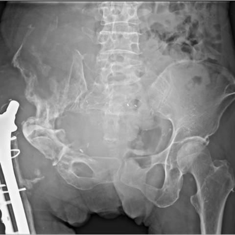
Frontal pelvic radiograph (2013) (Figure 1) showed THA with cortical irregularity and sclerosis of right iliac bone at the acetabular cup with adjacent hematoma.
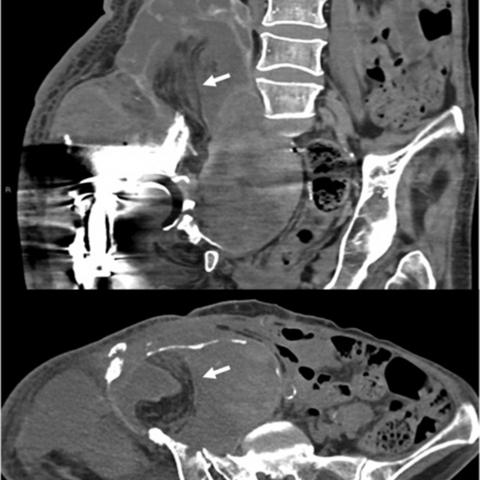
Axial computed tomography (CT) (2017) (Figure 2) showed a hematoma with bony erosion at the right iliac wing.
Contrast CT femur (2020) (Figure 3) demonstrated active arterial sub-capsular haemorrhage with bone erosion and remodelling. Digital subtraction angiography of the right internal iliac artery (2020) (Figure 4) showed extravasation into the hematoma.
Magnetic resonance imaging (MRI), axial T2-weighted image (Figure 5) showed susceptibility artefacts within an expanding aggressive hematoma.
Frontal pelvic radiograph (2021) (Figure 6) showed explantation of right acetabular cup with deformity of right ilium, acetabulum, and pubic rami and calcified rim of hematoma near the medial aspect of the proximal femur.
Coronal and Axial CT (2021) (Figure 7) demonstrated the presence of a pedicled musculocutaneous anterolateral thigh flap in the region of the right ilium.
Discussion
Background
Post-surgical hematoma that persists and enlarges over time is referred to as chronic expanding hematoma (CEH) [1]. Patients with bleeding diathesis and anticoagulant therapy have a higher risk of developing hematomas. Although not completely understood, it is hypothesized that the pathogenesis of CEH is due to fibroblastic reaction in response to the development of the fibrin matrix and cellular breakdown products of haemoglobin and platelets within the hematoma. This activates the inflammatory process, increases vascular permeability, and instigates bleeding from the fragile and dilated capillaries lining the internal cavity of the hematoma [2, 3]. Over time this results in hematoma expansion and in our case, eventual adjacent iliac wing osteolysis.
Clinical perspective
CEH may manifest as anaemia due to continuing blood loss, compressive mass effect over adjacent structures such as neurovascular bundle and ureter, and aggressive behavior such as osteolysis and bone remodelling. Our patient presented with severe pain and flexion deformity at the hip with inability to ambulate owing to aggressive osteolysis and bone destruction.
Imaging perspective
The usual CT appearance of an acute/ subacute hematoma is a hyperdense lesion (attenuation value 40-75 HU) with a smooth margin. Chronic hematoma is hypodense on CT due to old blood products and may demonstrate a pseudocapsule or a fibrous capsule that demonstrates peripheral enhancement related to new capillaries and granulation tissue [4].
MRI demonstrates internal low, intermediate, and high signals on T1 and T2-weighted imaging related to blood products of varying ages. Enhancement of the irregular pseudocapsule, granulation tissue, and new capillaries can make it challenging to differentiate CEH from a soft tissue sarcoma [5, 6]. Pseudocapsule with low signal intensity on T1- and T2-weighted imaging is also characteristic of CEH [7].
DSA shows active arterial blush in multiple feeding vessels as seen in our patient.
Histologically, CEH appears as a pseudocystic blood-filled lesion with a pseudocapsule composed of fibrous tissue with hemosiderin deposits and iron-laden macrophages [1, 3].
Outcome
The primary treatment of CEH is embolization. Prophylactic fixations are performed in areas of bone erosion and weakening. Complete surgical excision, including resection of the pseudocapsule is considered the gold standard treatment as it allows obtainment of the definitive histologic diagnosis. Incomplete removal may lead to recurrence [8].
Teaching point
CEH is a rare diagnosis with refractory bleeding. Differentiating from malignant tumours can be challenging.
Written informed patient consent for publication has been obtained.
Differential Diagnosis List
Final Diagnosis
Aggressive osteolysis by chronic expanding hematoma
Liscense
This work is licensed under a Creative Commons Attribution-NonCommercial-ShareAlike 4.0 International License.
Figures
Pelvic Radiograph

CT Pelvis

CT Femur

DSA angiography of the right internal iliac artery

MRI Pelvis

Pelvic Radiograph
CT Pelvis
Medical Imaging Analysis Report
I. Imaging Findings
Based on the provided X-ray, CT, MRI, and Digital Subtraction Angiography (DSA) images, the main findings are as follows:
- X-ray: A large abnormal density with relatively well-defined borders is observed near the right iliac bone. It is accompanied by local bone defect, bony destruction, and notable osteopenia. The right hip prosthesis remains in place, but an abnormal radiolucent area around it suggests possible soft tissue or hematoma-related lesions.
- CT Scan: A sizable low-density region is visible on the right side of the pelvis, showing a mixed density internally. Some parts at the boundary show enhancement, indicating fibrous or “pseudo-capsule” formation. Local erosive bone destruction in the right iliac bone is extensive.
- MRI: Mixed signal intensity is seen on T1- and T2-weighted images, suggesting both chronic and active bleeding components. A thick pseudo-capsule is visible around the lesion, which shows a ring of low signal intensity on T1 and T2 sequences. After contrast enhancement, the capsule and intralesional neovascular structures exhibit marked enhancement.
- DSA: Multiple feeding arteries demonstrate arterial blush during angiography, indicating that numerous feeding vessels are involved in the lesion and there is evidence of ongoing bleeding.
II. Potential Diagnoses
Based on the imaging features and the patient’s history of long-term repeated bleeding, the following diagnoses should be considered:
- Chronic Expanding Hematoma (CEH): This condition can result from postoperative or recurrent bleeding, leading to a chronic hematoma with a fibrous capsule. Radiologically, it features a “pseudo-capsule” enhancement and mixed density (or signal), and can bleed repeatedly.
- Soft Tissue Sarcoma: Various sarcomas can present as irregular soft tissue masses, with capsule-like enhancement or necrotic areas. They often have a rich blood supply and show more pronounced infiltrative growth, typically enlarging quickly or causing pain.
- Other Hemorrhagic or Vascular Lesions: Such as arteriovenous malformations or pseudoaneurysms formed after arterial aneurysm rupture. On DSA, these typically display more characteristic arteriovenous shunting or sac-like arterial aneurysms.
III. Final Diagnosis
Taking into account the patient’s advanced age, repeated bleeding episodes following hip surgery, the imaging findings of a “pseudo-capsule,” mixed density/signal, arterial bleeding signs, and pathological evidence of fibrous capsule and chronic hematoma, the most likely diagnosis is:
Chronic Expanding Hematoma (CEH)
If further confirmation is necessary, surgical excision and histological examination can be considered to exclude malignancies and prevent recurrent bleeding.
IV. Treatment Plan and Rehabilitation
1. Treatment Strategy
- Interventional Embolization: For recurrent or active bleeding, angiography followed by embolization can be performed first to control bleeding and alleviate acute symptoms.
- Surgical Intervention: Encompasses thorough removal of the hematoma and its fibrous capsule. Bone defect repair or orthopedic reconstructive procedures may be required if necessary. Complete removal reduces the risk of recurrence.
- Prevention of Complications: Pay attention to postoperative infection risks, further bleeding, and other complications. For elderly patients with multiple surgeries, preoperative assessments of cardiac and pulmonary function, as well as coagulation status, are particularly important.
- Supportive Care: Correct anemia, maintain adequate nutrition, closely monitor coagulation profiles, and adjust anticoagulant or antiplatelet therapy when needed to reduce intraoperative and postoperative bleeding risks.
2. Rehabilitation and Exercise Prescription
Due to the long disease course, multiple surgeries, and severe bone and soft tissue damage, an individualized and gradual rehabilitation program is recommended:
- Early Rehabilitation (Phase I):
• Objective: Promote wound healing and maintain basic functional mobility
• Methods: In-bed active and passive joint exercises, avoiding excessive weight-bearing; basic respiratory training to prevent pulmonary complications from prolonged bed rest
• Frequency and Duration: 1–2 times daily, 5–10 minutes each session, low intensity, focusing on slow movements - Mid-Stage Rehabilitation (Phase II):
• Objective: Gradually restore muscle strength and coordination, preventing muscle atrophy
• Methods: Under professional guidance, strengthen core and peripheral muscles using resistance bands or resistance training; if feasible, and with proper assistive devices or prosthetics, initiate standing balance exercises
• Frequency and Duration: 3–4 times weekly, 15–20 minutes per session, adjusted according to stamina and surgical recovery; intensity is increased gradually while avoiding strenuous movements - Late Rehabilitation (Phase III):
• Objective: Improve overall function and independence in daily activities
• Methods: Incorporate varied aerobic exercises (e.g., modified cycling, swimming, or other low-impact activities) and continue strengthening exercises to improve muscle power and joint flexibility
• Frequency and Duration: 3–5 times weekly, 20–30 minutes per session, progressively increasing the intensity and duration while keeping the heart rate within safe limits
Throughout the rehabilitation period, closely monitor the patient’s local condition and overall status. Adjust or temporarily suspend the program if necessary to ensure safety.
Disclaimer:
This report is a reference analysis based on existing historical and imaging data. It does not replace in-person consultations or guidance from a professional physician. The patient’s specific treatment and rehabilitation plan should be determined by professional medical evaluation and tailored to the clinical situation.
Human Doctor Final Diagnosis
Aggressive osteolysis by chronic expanding hematoma