


Complaints of prostatism and pain in the right hip. History of renal carcinoma which was treated by nephrectomy 8 years ago and Paget’s disease of the right hemipelvis and proximal right femur known for 20 years.
The patient complained of prostatism and pain in the right hip. His medical history revealed a renal carcinoma which was treated by nephrectomy 8 years before and Paget’s disease of the right hemipelvis and proximal right femur known for 20 years. Plain radiograph and CT scan were performed and showed Paget’s changes at the pelvis and an osteolytic lesion proximal in the right femur. Subsequently MRI and needle biopsy under CT guidance were carried out. Histology after CT guided biopsy of the right trochanteric region showed a metastasis of renal cell carcinoma.
Neoplastic involvement in Paget’s disease is possible and includes sarcomatous transformation, giant cell tumor transformation, superimposed metastatic disease (breast, lung, kidney, prostate and colon), plasma cell myeloma or lymphoma. There are no studies reporting the incidence of metastases compared to sarcomatous degeneration. Moreover, there are conflicting reports whether metastases occur more or less frequently than metastases in normal bone. Increased local blood flow in Paget’s disease could be responsible for increased metastatic disease. Both sarcomatous degeneration and metastatic lesions may be responsible for increased local pain and soft tissue swelling. Radiologically osteolytic lesions within a pagetic bone and/or a soft tissue mass can be found. The lytic lesions are best demonstrated on conventional radiographs and CT, whereas soft tissue extension is best visualized on MRI. Needle biopsy with histological study of the specimen is mandatory to confirm the presumed diagnosis.
Metastasis of renal cell carcinoma

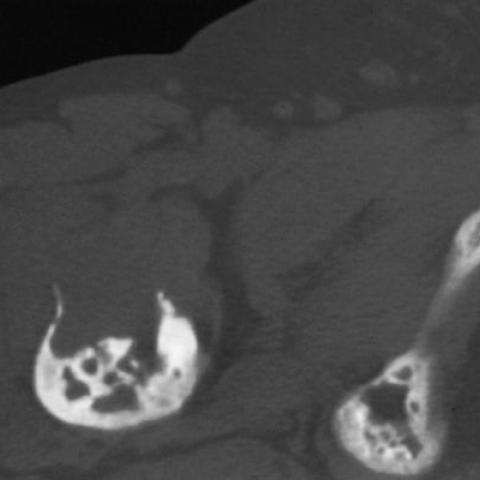


1. X-ray and CT demonstrate significant thickening of the right iliac bone and proximal femur, with disordered trabecular patterns and mixed densities, consistent with the known Paget disease lesions in the right pelvis and femur.
2. Focal osteolytic areas are observed, with irregular cortical bone and relatively unclear boundaries.
3. MRI shows abnormal signals in the right proximal femur and pelvic region, with a soft-tissue mass-like lesion, indicating that the disease has extended into extraosseous soft tissue.
4. Overall, imaging findings suggest the possibility of a new or superimposed lesion on the existing Paget disease. Considering the patient’s clinical symptoms (increased right hip pain, prostate issues, etc.), there is a high suspicion of malignant transformation (e.g., sarcoma) or metastatic disease.
1. Sarcomatous transformation of Paget’s disease: Long-standing Paget’s disease may develop malignant tumors such as osteosarcoma or chondrosarcoma. Radiologically, previously thickened bone may exhibit local osteolytic lesions or soft-tissue masses.
2. Metastatic lesions:
3. Other rare malignancies: Such as multiple myeloma or lymphoma, which should also be considered in the differential diagnosis. However, based on the patient’s clinical status and history, these are less likely.
Given the suspicious soft-tissue mass and osteolytic changes noted on imaging, along with worsening pain, a high suspicion arises for malignant transformation (sarcoma) or metastatic lesions on the basis of Paget’s disease. The most likely current diagnosis is a metastatic lesion or sarcomatous lesion, pending histopathological biopsy confirmation.
1. Treatment Strategy
2. Rehabilitation / Exercise Prescription
During the diagnostic and treatment phase, the patient should engage in gradual and supervised activity under the guidance of physicians and rehabilitation therapists. Key points include:
[Disclaimer]
This report is based on the provided clinical history and imaging data for reference only, and cannot replace face-to-face consultations or professional medical advice. The final diagnosis and treatment plan should be formulated by specialists from the relevant departments, taking into account the patient’s specific condition.
Metastasis of renal cell carcinoma