Staphylococcus aureus spondylitis: MR and helical CT findings


Clinical History
Progressive low-back pain and elevated erythrocyte sedimentation rate in HIV-positive drug addict.
Imaging Findings
The patient, an HIV-positive drug addict, presented with progressive low-back pain, throbbing at rest and exacerbated by motion, general malaise, anorexia and limited weight loss. At physical examination spinal tenderness and rigidity were observed. Blood examinations were within the normal range apart from the erythrocyte sedimentation rate, which was elevated.
On magnetic resonance imaging the lower half of the L2 vertebral body showed a focus of signal abnormality that on T2-weighted images was characterised by a "target-type" appeareance (a high signal intensity lesion with low signal intensity peripherally and in the centre). On post-contrast T1-weighted images the lesion showed a thin peripheral rim of enhancement. The lesion was associated with bone marrow oedema and lower end-plate lysis. The L2/L3 intervertebral disc was slightly decreased in height and showed constantly a low signal intensity on both T1- and T2-weighted images in the absence of significant enhancement. Prominent epidural soft tissue was seen on the anterior and posterior left lateral aspect of the spinal canal causing slight impingement and dislocation of the thecal sac. On the CT image a higher attenuation of the spongiform bone trabeculae surrounding the lesion was seen. A biopsy of the lesion and a culture of the bioptic material were performed and the definitive diagnosis was: Staphylococcus aureus spondylitis.
Discussion
The key feature in this case is the observation of a preminently pure infective spondylitis rather than the more frequently observed spondylodiscitis. In intravenous drug abusers clinical diagnosis of spondylitis may be difficult due to extraspinal abnormalities overshadowing the vertebral alterations. Diagnosis may be difficult also in patients with operated disc herniation, in young children and in paraplegic or para-quadriplegic patients (1). With diffusion of MRI into everyday clinical practice the reported frequency of vertebral osteomyelitis and disc space infection has risen dramatically from less than 1% to 2-4% of all cases (1). In adults haematogenous spread of infection to the subchondral regions of the vertebral endplate adjacent to the intervertebral disc is followed by endplate perforation, extension to the contiguous vertebral body and narrowing of the interposed intervertebral disc (1). Infection may then extend to the ventral and/or dorsal surface of the vertebra in a subligamentous fashion and eventually to the surrounding soft tissues. After a variable period (4 to 10 weeks) a reactive osteosclerotic response of the surrounding bone is observed (1).
In this case narrowing of the intervertebral disc in the absence of signal abnormality and postcontrast enhancement could be generically ascribed to chronic degenerative disc disease thus misleading the diagnosis. On the other hand a bony fragment within the lesion was highly suggestive of an osteomyelitic process as well as the accompanying osteosclerotic response of the spongiform bone. This is reported to be a helpful sign in differentiating pyogenic from tuberculous spondylitis (2,3). Tuberculous and other granulomatous spondylitis (syphilis, sarcoidosis and fungal disorders) are usually characterised by a slowly progressive vertebral lesion, preservation of the intervertebral disc, subligamentous spread with erosion of the anterior vertebral margins, large prevertebral soft tissue abscesses usually involving the psoas muscles and absence of severe bony sclerosis (eburnation). On the other hand pyogenic spondylitis is more frequently characterised by a rapid loss of the intervertebral disc height (usually associated with high T2 signal hyperintensity and enhancement on MR), posterior extension of the process (epidural abscess) and marked early reactive bony eburnation. The latter is usually missed on MR and very well appreciated on CT scans. Several other processes may produce similar abnormalities on MRI.
Acute cartilagineous node formation (Schmorl's node) is usually associated with a decrease in intervertebral disc height and bony sclerosis; however in this case the acute junction angles of the lesion with the vertebral endplate stood for a lesion originating within the vertebral body and not extending from the intervertebral disc.
Primary (focal and systemic) or metastatic neoplasias should also be included in differential diagnosis. Certain tumours such as plasma cell myeloma, lymphoma, chordoma, giant osteoblastoma and even skeletal metastases can extend around the intervertebral disc to involve the neighbouring soft tissues. The combination of focal or widespread lysis or sclerosis of a vertebral body and an intact intervertebral disc is much more characteristic of tumour than infection (4).
Differential Diagnosis List
Final Diagnosis
Staphylococcus aureus spondylitis
Liscense
Figures
Unenhanced sagittal SE T1-weighted images (TR/TE/NEX:500/15/2)

Enhanced sagittal SE T1-weighted images (TR/TE/NEX:500/15/2)


Enhanced axial SE T1-weighted images (TR/TE/NEX:500/15/2) at level of the L2 lower end-plate


Sagittal TSE T2-weighted image (TR/TE/ETL/NEX:2200/120/16/4)

L2 lower end-plate scan during needle-biopsy

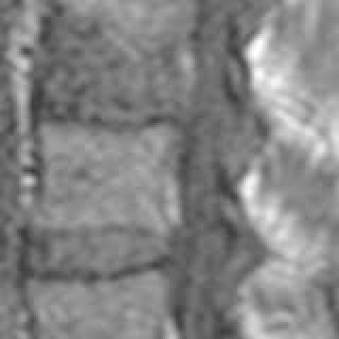
Sagittal reconstruction with high spatial resolution algorithm and bone window settings


1. Imaging Findings
Based on the provided lumbar spine MRI and CT images, the following main features are observed:
• The lesion is primarily located in one of the lumbar vertebrae (initially appearing in the mid-lumbar region), showing an abnormal signal within the vertebral body, with bone destruction or defect along the anterior border of the vertebral body.
• The adjacent intervertebral disc space is narrowed to varying degrees, but no significant high signal or enhancement suggestive of infection is seen on MRI. This differs from the common disc-involving presentation (“discitis” or purulent spondylodiscitis).
• Within the vertebral body, hyperdense areas resembling free bone fragments or sequestra are visible (more clearly on CT), surrounded by prominent sclerosis, suggesting a reactive bone change following infection.
• A possible paravertebral or intraspinal soft tissue shadow is noted, raising the suspicion of soft tissue involvement or abscess formation (requiring contrast-enhanced scans and correlation with clinical symptoms for confirmation).
2. Potential Diagnoses
Considering the above imaging features, along with the patient’s background of HIV positivity, intravenous drug use, and elevated ESR, the following differential diagnoses are possible:
- Purulent Vertebral Osteomyelitis (Spinal Infection): Often caused by bacteria spreading via the bloodstream to the vertebral body or intervertebral disc. Intravenous drug users and immunocompromised individuals are at higher risk. Typical manifestations include vertebral destruction, sclerosis, and/or paravertebral soft tissue abscess. Adjacent intervertebral discs are frequently involved, but predominant vertebral lesions can also occur.
- Tuberculous Spondylitis (Spinal Tuberculosis): Usually has a chronic course, commonly presenting with vertebral destruction and paravertebral or psoas abscesses. Intervertebral discs are often involved at a later stage, and sclerosis is generally less pronounced than in purulent infections.
- Other Granulomatous or Fungal Infections: For example, fungal, syphilitic, or candidal infections, which can also lead to vertebral destruction. Their progression is slower and resembles tuberculosis, typically with less sclerosis.
- Neoplastic Lesions: Such as spinal metastases, lymphoma, or myeloma, which can produce osteoblastic or osteolytic destruction and may involve surrounding soft tissues. Generally, the intervertebral disc is relatively preserved, and neoplastic lesions may show distinct enhancement patterns on CT or MRI.
3. Final Diagnosis
Given the patient’s immunocompromised status (HIV-positive), history of intravenous drug abuse, elevated ESR, and imaging findings of localized vertebral destruction, bone sclerosis, and suspected sequestrum-like changes, the most likely diagnosis is: Purulent Vertebral Osteomyelitis (Pyogenic Spondylitis).
The most common causative organisms include Staphylococcus aureus or other bacteria (e.g., Pseudomonas). Confirmation requires serological tests or image-guided biopsy and culture.
4. Treatment Plan and Rehabilitation
Treatment Strategy:
• Antimicrobial Therapy: Empirical broad-spectrum antibiotics, adjusted according to culture and sensitivity results. The course of treatment is usually long, at least 6–8 weeks.
• Bracing or Bed Rest: During the acute phase, a lumbar brace can be used to reduce weight bearing. Strict bed rest requires regular repositioning to prevent pressure sores.
• Surgical Intervention: In cases of spinal instability, large abscesses causing compression, or failure of medical therapy, surgical debridement and spinal fusion may be considered to stabilize the spine and promote healing.
Rehabilitation / Exercise Prescription (FITT-VP Principle)
After controlling the acute infection, individualized and gradually progressive rehabilitation is recommended, following these principles:
1. Frequency (F): Start with 2–3 sessions per week; increase to 3–5 sessions weekly based on tolerance.
2. Intensity (I): Begin with low intensity and gradually increase as tolerated. Aim for a level where the patient experiences mild fatigue.
3. Time (T): Start with 10–15 minutes per session and gradually extend to 30 minutes; splitting sessions is also possible.
4. Type (T): Emphasize safe, low-impact exercises, such as core strengthening in the supine position (bridges, leg lifts), and moderate swimming or water-based activities. Avoid heavy lifting or extreme lumbar flexion/extension exercises.
5. Progression (P): As pain and inflammation improve, gradually increase range of motion and load, for instance moderate walking or lower limb strengthening, under the guidance of a specialist or rehabilitation therapist.
Monitor spinal stability and the patient’s overall condition (including HIV control and nutritional status). If severe pain or neurological symptoms worsen, prompt re-evaluation is necessary.
5. Disclaimer
This report is based solely on the provided imaging and partial clinical information, and cannot replace an in-person consultation or professional medical opinion. Specific treatment plans must be determined by a specialist after a comprehensive review of the patient’s complete clinical data and subsequent examination results.
Human Doctor Final Diagnosis
Staphylococcus aureus spondylitis