Fluorosis: CT and MR imaging findings


Clinical History
The patient was admitted with the signs and symptoms of paralysis of the arms and legs and diffuse bone and low-back pain.
Imaging Findings
The patient was admitted with the signs and symptoms of paralysis of the arms and legs and diffuse bone and low-back pain. She also complained of forgetfulness of 2 years' duration and loss of concentration when performing detailed functions in daily life. Laboratory results were within the normal limits.
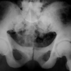
Plain radiography of the skeletal system demonstrated sclerotic changes in both the axial and appendicular skeleton (Fig. 1). On computed tomography (CT), the bones showed prominent cortical thickening and increased density with irregular contours. Bony excrescences were detected in both the pelvic bones and the lower extremities. The sacroiliac joints were narrow(Fig. 2).
Magnetic resonance imaging (MRI) showed prominent signal losses in all bones on spin-echo T1- and gradient-echo T2*-weighted images. On thoraco-lumbar MRI, the vertebrae and sternum showed prominent hypointense signals on both T1- and T2*-weighted images. The intervertebral disc spaces were decreased and multiple prolapses were demonstrated (Fig. 3). The medullary canal was narrowed by bony excrescences and by the ossified posterior longitudinal ligament. Axial MRI sections through the pelvic bones showed hypointense signals on both T1- and T2*-weighted images (Fig. 4). Bony excrescences were also demonstrated on MRI. On cranial MRI, the brain showed atrophic changes, particularly prominent bilaterally at the perislyvian areas, despite the patient's young age (Fig. 5).
Discussion
Fluorosis is related to high concentrations of fluoride present in drinking water, surrounding soil or produced by coal burning. It is endemic in certain parts of the world, including the eastern part of Turkey. Its effects on the skeletal system and teeth are well known. Fluoride directly stimulates bone formation in vivo and fluoroapatite formation (1). Involvement of the axial skeleton is characteristic. Fluoride is retained in the bones and induces hardening of all the bones, including the spine. Increased trabecular condensation creates an extremely radiodense appearance throughout the thorax, vertebral column and pelvis. The skull and tubular bones are generally relatively spared. However, in this case the changes in tubular bones were also very prominent. This can be attributed to high exposure or a long duration of exposure to fluoride (1).
Vertebral osteophytosis may be very prominent and can encroach on the spinal canal and vertebral foramina. Thus neurological symptoms are frequently seen, as in this case.
Bony excrescences develop at the sites of ligamentous insertions (iliac crests, ischial tuberosities and inferior margins of ribs). In the appendicular skeleton osteopaenia with or without recovery lines may be an early finding. Osteophytosis, ligament calcification and periosteal thickening can also be seen.
The common findings of osteosclerosis, osteophytosis, ligamentous calcification and periostitis can also be seen in many other diseases. When these findings are detected in a patient coming from a non-endemic region, the following differential diagnoses should be considered: skeletal metastasis, especially prostate metastasis; myelofibrosis; mastocytosis; renal osteodystrophy; Paget's disease; pachydermoperiostitis; and diffuse skeletal hyperostosis. The demonstration of a primary malignancy in the prostate and the age of the patient can provide important clues in prostate carcinoma. Metastatic disease is less generalised, less symmetric, and most frequently involves the axial skeleton. Myelofibrosis is seen in middle-aged or elderly patients. It is characterised by bone marrow fibrosis and splenomegaly (2). Mastocytosis may produce local or diffuse sclerosis associated with hepatosplenomegaly. Paget's disease causes a characteristic coarsened trabecular pattern (3). Pachydermoperiostosis is easily differentiated by digital clubbing, skin changes, enlargement of the paranasal sinuses, and the predominant feature of periostitis extending to the epiphysis (4). Osteosclerosis is a well-known feature of renal osteodystrophy. It predominates in the axial skeleton and is most prominent in the superior and inferior portions of the vertebral bodies (rugger-jersey spine). In diffuse idiopathic skeletal hyperosteosis, ligament calcification and ossification is present along the anterolateral portion of at least four contiguous vertebral bodies. Sclerotic changes, like dripping candle wax in the medial portion of the tibia, can mimic melorheostosis, as in this case. But in melorheostosis this sign especially affects the outer rather than inner surface of the affected limb. In contrast to fluorosis, melorheostosis most commonly affects the tubular bones (5).
Fluorosis can affect many other body systems, including the respiratory, urinary and central nervous systems. The blood-brain barrier fails to exclude fluoride from nervous tissue and, consequently, this ion accumulates in the brain. In affected patients many kinds of neurological symptoms have been reported, such as partial paralysis of the arms and legs, headache, spasticity in the extremities, visual disturbance and mental retardation. In this case, despite the patient's young age, the brain showed significant atrophic changes.
Differential Diagnosis List
Final Diagnosis
Fluorosis
Liscense
Figures
Plain radiography appearance of fluorosis


CT findings of fluorosis


MR imaging appearances of the thoracic spine


MR imaging appearance of pelvic bones


MR imaging appearance of brain

Medical Analysis Report
1. Imaging Findings
Based on the provided X-ray, CT, and MRI images, the following main features are observed:
- Increased Bone Density: In the pelvis, vertebral bodies, and other locations, the trabeculae appear denser, presenting a pronounced high-density pattern overall.
- Osteophyte Formation at Vertebral Margins: Multiple vertebral segments show noticeable osteophytes, some protruding into the spinal canal or intervertebral foramen, potentially causing nerve compression.
- Enthesopathic Changes or Sclerosis: Abnormal proliferations or sclerotic changes may be observed at certain ligamentous or tendon attachment sites, such as the iliac bone and ischial tuberosity.
- Local Soft Tissue: No obvious large soft tissue masses are noted, but in some areas, osteophytes or ligament calcification lead to narrowed soft tissue spaces.
- Brain Changes: MRI indicates signs of brain atrophy, which may be related to the clinical symptoms (weakness in the limbs, possible neurological involvement, etc.).
2. Potential Diagnoses
Combining the patient’s profile (35-year-old female), clinical manifestations (quadriplegia, bone pain), and imaging features (increased bone density, osteophytes, and signs of nerve compression), the differential diagnoses may include:
- Skeletal Fluorosis (Fluoride Poisoning): Common in high-fluoride environments (water, air, or prolonged exposure to fluoride substances). Characterized by widespread bone density increase, notable vertebral and pelvic sclerosis, osteophyte formation causing nerve compression, and other system-related symptoms.
- Diffuse Idiopathic Skeletal Hyperostosis (DISH): Primarily involves ossification of ligaments and tendon attachments with relatively symmetrical anterior vertebral body thickening. However, widespread increase in bone density is not always present, and clinical symptoms may differ slightly from this case.
- Sclerosing Bone Diseases (e.g., Myelofibrosis, Mastocytosis, Paget’s Disease, etc.): These conditions can also cause bone sclerosis, but they are often accompanied by other systemic features (e.g., splenomegaly, hepatomegaly, or characteristic bone changes), distinguishing them from this case.
- Metastatic Bone Disease: For instance, osteoblastic metastases from prostate cancer typically present sclerotic lesions, but they are rare in females and often appear asymmetrical.
- Other Rare Sclerosing Bone Disorders: Such as melorheostosis, which often involves tubular bones primarily, and its distribution typically does not match the widespread pattern seen in this case.
3. Final Diagnosis
Taking into account the patient’s environmental background (possibly from or long-term residence in a high-fluoride area), clinical manifestations (bone pain, limb paralysis reflecting neurological involvement), and imaging features (widespread bone sclerosis, dense vertebrae and pelvic bones, and brain atrophy suggesting neurological implications), the most likely diagnosis is:
Skeletal Fluorosis (Fluoride Poisoning).
If fluorine levels or bone metabolism indices are not yet confirmed, measuring serum and urinary fluoride levels, along with other bone metabolism markers, can help verify the diagnosis.
4. Treatment and Rehabilitation Plan
4.1 Treatment Strategies
- Eliminate or Minimize Fluoride Exposure: Such measures include switching the water source, avoiding exposure to fluoride-containing coal combustion, and enhancing environmental protection.
- Symptomatic Supportive Treatment: For significant pain, pain relievers, non-steroidal anti-inflammatory drugs, or auxiliary physical therapy may be considered. Supplementation with calcium and vitamin D may be used to improve bone quality.
- Nerve Compression Management: In cases where severe spinal cord or nerve compression is caused by osteophytes, neurosurgical or orthopedic interventions (e.g., surgical decompression) may be an option.
- Other Supportive Therapies: If there is evidence of brain dysfunction or marked neurological injury, coordinated rehabilitation with neurology or physical medicine may be required.
4.2 Rehabilitation / Exercise Prescription Recommendations
During rehabilitation, considerations must be given to bone pain, altered bone structure, and neurological involvement. An individualized and gradual approach to exercise is advised:
- Early Stage (Acute / Painful Phase):
- Focus on passive joint movements and gentle active exercises, avoiding heavy weight-bearing or extreme range of motion.
- Perform low-intensity exercises 2-3 times a day, each session about 10 minutes, such as bedside leg lifts, ankle pump exercises, and gentle stretching.
- Intermediate Stage (Functional Recovery Phase):
- Gradually introduce muscle strengthening exercises, such as resistance band workouts and light resistance training (e.g., seated leg lifts, wall squats).
- Depending on the patient’s tolerance, each session can last 15-20 minutes, 3-4 days a week, while monitoring pain and fatigue levels.
- Late Stage (Strengthening Phase):
- Emphasize core and lower limb strength to improve stability and balance, e.g., standing balance drills, slow walking, or low-intensity elliptical training.
- Each session can be 20-30 minutes, 3-5 days a week, gradually increasing exercise load but avoiding sudden impacts or twisting injuries.
Throughout the entire rehabilitation process, pay special attention to:
- Monitoring bone pain, lower limb numbness, and neurological function. Seek medical advice if symptoms worsen significantly.
- Controlling exercise intensity to prevent secondary injuries due to compromised bone or nerve integrity.
- Using assistive devices (such as lumbar supports or braces) if necessary to protect spinal stability.
5. Disclaimer
This report is a reference medical analysis based on the provided medical history and imaging data. It cannot replace in-person clinical diagnosis and professional medical treatment. For final decisions regarding diagnosis and treatment, please consider clinical examinations, laboratory tests, and the evaluation of specialized physicians.
Human Doctor Final Diagnosis
Fluorosis