Ivory vertebra: non-Hodgkin's lymphoma


Clinical History
Back pain, discomfort and loss of sensation along both legs for a few months.
Imaging Findings
The patient presented with back pain, discomfort and loss of sensation along both legs for a few months. Plain films of the lumbar spine were performed which showed a dense third lumbar vertebra, giving an appearance of an ivory vertebra.
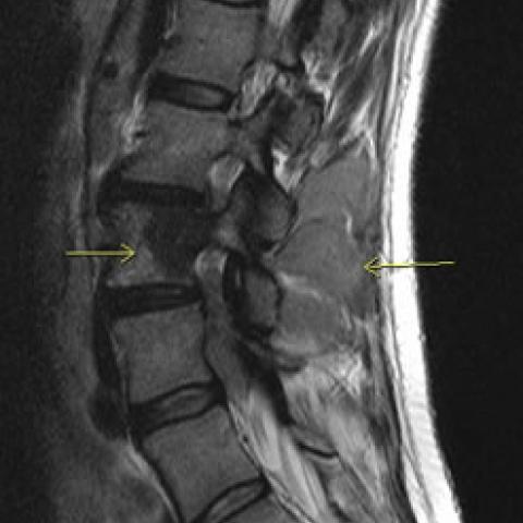
In view of the patient's symptoms, an MRI of the lumbar spine was performed. This showed an abnormal marrow signal of the L2 and L3 vertebrae associated with involvement of the posterior elements. An abnormal soft tissue mass was noted in the spinal canal, also extending to involve the right psoas muscle and the right erector spinae muscle. A CT-guided biopsy of the soft tissue mass was performed at multiple levels. Pathological diagnosis was of a low-grade non-Hodgkin's lymphoma. The patient was referred for treatment.
Discussion
Solitary dense vertebrae, as well as in lymphoma, are also seen in osteoblastic metastases (prostate/breast), haemangioma (coarse trabecular pattern, vertical in orientation), Paget's disease (tends to involve the posterior elements as well as displaying expansion of the body), osteopetrosis, sickle cell anaemia, fluorosis, systemic mastocytosis, tuberculous infection, metastatic carcinoid tumour, osetoblastoma, osteosarcoma and even primary Ewing's sarcoma
Skeletal non-Hodgkin's lymphoma is more commonly a manifestation of a diffuse disease than a primary tumour. It is seen in 10-20% of adults and 20-30% of children with the disease. Disseminated disease is related to haematogenous spread and involves the spine, pelvis, skull and ribs. Primary lymphoma of the bones can occur at any age and is more common in males. Systemic symptoms are usually absent. Local swelling and pain is common. Lesions tend to predominate in the appendicular skeleton, especially in the lower extremities. Pathological fractures are common.
Histologically the most common types are histocytic lymphoma or poorly differentiated or undifferentiated lymphomas. Prognosis depends on the histological type.
Differential Diagnosis List
Final Diagnosis
Ivory vertebra: non-Hodgkin's lymphoma
Liscense
Figures
AP/lateral view of the lumbar spine


MRI of the lumbar spine

CT lumbar spine: prone


Imaging Findings
Based on the provided X-ray, CT, and MRI images, one lumbar vertebral body appears to have increased density (i.e., “sclerotic” or higher-density changes), with abnormal local bony structure and signal alterations compared to normal bone. The vertebral margins remain relatively intact, but there is a degree of substantial lesion involvement within the vertebral body. On MRI, the lesion shows abnormal signals on T1- and T2-weighted sequences; after contrast enhancement, the lesion area demonstrates enhancement, along with signs of anterior epidural or paravertebral soft tissue involvement in some cases.
These findings also suggest that the lesion may cause some degree of spinal canal, nerve root, or soft tissue compression, correlating with the patient’s clinical symptoms (lower back pain, bilateral leg sensory abnormalities, and possible nerve root compression symptoms).
Potential Diagnoses
Based on the patient’s age, clinical symptoms, and imaging findings, the following diagnoses or differential diagnoses are worthy of consideration:
- Primary or Secondary Bone Lymphoma (Non-Hodgkin’s Lymphoma): May present with density or signal abnormalities within the vertebral body, commonly seen in older adults. It can be a primary bone lymphoma or part of a systemic lymphoma involving the spine.
- Metastatic Vertebral Tumor (e.g., breast cancer): In a female patient, if there is a history of breast cancer or other malignancies, metastatic spinal lesions should be strongly suspected. Sclerotic-type metastases may appear as “dense vertebrae.”
- Vertebral Hemangioma: Typically, imaging may show characteristic vertical striations (“honeycomb” or “polka-dot” appearance). It usually lacks a significant soft tissue mass, but must be distinguished from other sclerotic vertebral lesions.
- Paget’s Disease: Commonly presents with bone thickening and disorganized trabecular structure, possibly involving the posterior elements of the vertebra, accompanied by pronounced bone overgrowth and deformities.
Definitive diagnosis requires correlation with clinical history, laboratory tests (such as tumor markers, LDH, complete blood count), and necessary pathological investigations.
Final Diagnosis
Taking into account a 60-year-old female, recent months of lower back pain and bilateral lower limb sensory disturbances, imaging findings of a solitary vertebral body with increased density and possible local soft tissue involvement, the most likely diagnosis is Non-Hodgkin’s Lymphoma (NHL) of the Bone. However, other lesions like metastatic disease must be ruled out. Further enhanced imaging studies or vertebral biopsy are needed to confirm the pathological diagnosis.
Treatment Plan and Rehabilitation
1. Treatment Strategies:
- Chemotherapy and/or Radiotherapy: If the diagnosis of bone lymphoma is confirmed, chemotherapy and radiotherapy are the mainstays of treatment. Specific regimens should be determined in consultation with hematology or oncology specialists.
- Surgical Intervention: If there is significant spinal cord or nerve root compression leading to neurological deficits or a high risk of pathological fracture, surgical decompression or stabilization may be considered to maintain spinal stability and relieve pain and neurological symptoms.
- Supportive Care: Includes pain management, nutritional support, correction of anemia, and effective control of underlying conditions to improve the overall condition of the patient.
2. Rehabilitation / Exercise Prescription:
- Principles: After initial treatment (chemotherapy, radiotherapy, or surgery), rehabilitative exercise should follow the FITT-VP principles (Frequency, Intensity, Time, Type, Volume, Progression). Activity should be gradually increased according to individual tolerance and safety.
-
Early Phase (Recovery Stage):
- Frequency: 2-3 times a week, with at least 1 day of rest in between sessions.
- Intensity: Light to moderate intensity (e.g., 5-10 minutes of level walking or mild stretching exercises).
- Time: Keep each exercise session under 20 minutes, possibly divided into segments.
- Type: Gentle aerobic exercises (such as level walking or stationary cycling) and core muscle strengthening (e.g., supine leg raises, pelvic lifts) performed in bed or on a mat.
-
Progressive Phase (Consolidation Stage):
- As tolerated and if the condition allows, increase the exercise volume or duration by 5-10% weekly.
- Moderately enhance core stability and lower limb strength training, such as seated light resistance exercises (using resistance bands or light dumbbells), avoiding heavy loads and excessive forward bending.
- Gradually extend the session duration up to around 30 minutes; if significant pain or fatigue occurs, reduce the exercise volume accordingly.
- Special Considerations: If bilateral leg numbness worsens, lower back pain intensifies, or if there is bowel or bladder dysfunction or abnormal blood counts, seek medical attention promptly and reassess the safety of exercise. Adjust or pause the exercise prescription as necessary.
Disclaimer
This report is a reference analysis based on the current imaging data and clinical information provided. It does not replace in-person medical consultations or professional medical advice. Patients should seek further diagnosis and treatment at a hospital and adjust their rehabilitation and exercise plan under the guidance of qualified healthcare professionals.
Human Doctor Final Diagnosis
Ivory vertebra: non-Hodgkin's lymphoma