


One year after complete AC joint dislocation she was experiencing significant symptoms of shoulder pain radiating into her neck and arm.
The patient sustained an injury to her right shoulder in a fall from a horse. On examination there was swelling and tenderness over the acromioclavicular (AC) joint and no evidence of any distal neurovascular deficit.
Radiographs showed a complete AC joint dislocation (Fig 1).
She was treated by immobilization in a collar and cuff for 3 weeks, followed by physiotherapy.
One year after the injury she was still experiencing significant symptoms of shoulder pain radiating into her neck and arm.
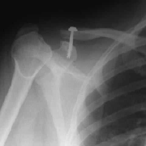
Radiographs taken at this stage showed erosive changes in the distal end of the clavicle (Fig 2). She was listed for AC joint reconstruction surgery. At the time of her surgery, two years after the initial injury, further radiographs showed that the osteolysis had progressed, with the lateral 2-3 cm of the clavicle having been resorbed. She was treated with screw fixation of the lateral end of the clavicle to the coracoid process and coracoclavicular ligament reconstruction (Fig 3). The screw was removed after nine months. After physiotherapy the shoulder was pain-free with a good range of movement.
Osteolysis of the lateral tip of the clavicle is a rare condition, having been first described by Werder in 1950 (1). It usually follows an injury to the AC joint, although the joint need not be subluxed in order to cause osteolysis. It has also been reported to occur without an acute injury in males who participate in weight-training (2) or use pneumatic tools (3). In post-traumatic cases characteristic resorption of the lateral end of the clavicle, with loss of subarticular cortical bone and cystic changes, although it may be seen on radiographs as early as two-and-a-half weeks after the injury (4), is usually seen within one to three months, and can progress for several years (5). Up to 2-3cm of the clavicle may be involved, and occasionally there is also ostelysis of the acromion. Clinical features are persisting shoulder pain and weakness, with swelling and tenderness over the AC joint. Because radiographs taken immediately following the injury usually show a normal clavicle, the diagnosis is often missed. Connective tissue disorders, hyperparathyroidism, infection, neoplastic disease and massive osteolysis are all important differential diagnoses which can give similar radiographic appearances. The pathological process involved remains unclear; ischaemia, an autonomic phenomenon or synovial overgrowth have all been suggested as possible mechanisms. Immobilization, resection of the lateral end of the clavicle, and AC joint reconstruction have all been proposed as treatment options.
Post-traumatic osteolysis of the clavicle


Based on the provided shoulder X-ray images (anterior-posterior and lateral/oblique views), the following key features are observed:
1. A noticeable anatomical abnormality at the acromioclavicular (AC) joint, especially an abnormal relationship between the lateral (distal) aspect of the clavicle and the articular surface of the acromion.
2. Evidence of irregular bone resorption at the distal clavicle, with partial loss of cortical continuity or blurred margins. Cystic or focal radiolucent areas are visible, suggesting bone destruction or resorption.
3. In some images, metal fixation (screws) is present near the distal clavicle, indicating the patient may have undergone previous joint reconstruction or internal fixation.
4. No obvious radiographic signs of swelling in the acromion or surrounding soft tissues, but there may still be chronic inflammatory or scar tissue changes in the area.
Taking into account the history of a complete AC joint dislocation one year ago and the current imaging findings, the potential diagnoses or differentials include:
1. Traumatic Osteolysis of the Distal Clavicle:
• Commonly occurs after AC joint injury, featuring progressive bone resorption of the distal clavicle and pain.
• Radiographic findings include bone destruction and thinning or defects of the distal clavicle cortex, possibly with changes in the joint space.
• Consistent with the clinical presentation of persistent shoulder pain following trauma.
2. Infectious Bone Destruction (e.g., osteomyelitis):
• Clinically associated with local redness, swelling, fever, or systemic signs of infection.
• Correlation with inflammatory markers (CRP, ESR, white blood cell count) and clinical features is necessary.
• In this case, no clear signs of infection have been noted, so it remains a differential diagnosis.
3. Metabolic Bone Disease (e.g., hyperparathyroidism):
• Can present with multiple areas of bone resorption, often involving other skeletal regions.
• Given that the changes are isolated to the AC joint and there is a known history of trauma, the likelihood is relatively low.
4. Tumor-Related Bone Destruction (benign or malignant):
• Typically includes progressively worsening pain or a palpable mass.
• In this case, based on the imaging features and traumatic history, a traumatic etiology is more likely.
Considering the patient’s prior AC joint dislocation, persistent shoulder pain, and distinctive radiographic signs of distal clavicle osteolysis, the most likely diagnosis is:
“Post-traumatic Osteolysis of the Distal Clavicle.”
Because of the significant and prolonged symptoms and the apparent presence of internal fixation (screws), the definitive diagnosis should be correlated with surgical records and laboratory tests (to exclude infection and metabolic diseases). If uncertainty remains or further differentiation is required, an MRI or, if necessary, a biopsy can be considered.
1. Treatment Strategies:
• Conservative Treatment: For milder symptoms, recommend restricting shoulder movement, using nonsteroidal anti-inflammatory drugs (NSAIDs) for pain relief, and localized physiotherapy.
• Joint Reconstruction or Surgical Treatment: For those who have had internal fixation but continue to experience significant symptoms or progressive osteolysis, a second surgical intervention may be warranted, such as partial distal clavicle resection or AC joint reconstruction.
• Rehabilitation and Functional Training: Whether surgery is performed or not, rehabilitation is critical to restoring shoulder range of motion, strength, and stability.
2. Rehabilitation/Exercise Prescription (FITT-VP Principle):
• Initial Phase (Postoperative or Acute Inflammatory Phase):
- Frequency: 1–2 short sessions per day according to pain tolerance.
- Intensity: Activities that do not cause pain or only slight sore sensations; avoid lifting heavy objects.
- Time: 10–15 minutes per session of gentle passive/assisted active exercises.
- Type: Passive joint mobilization, Range of Motion (ROM) exercises, gentle stretches for shoulder girdle musculature.
- Progression: Gradually increase ROM and incorporate strengthening exercises as tolerated.
• Intermediate Phase (Chronic Phase or Stable Postoperative Phase):
- Frequency: 3–4 times per week.
- Intensity: Light to moderate strength exercises, possibly with resistance bands or light dumbbells for shoulder abduction, adduction, and forward elevation.
- Time: 20–30 minutes per session.
- Type: Focus on strengthening the rotator cuff (supraspinatus, infraspinatus, teres minor, subscapularis) and scapular stabilizing muscles.
- Progression: Increase resistance or complexity as muscle strength improves.
• Late Phase (Functional Recovery and Return to Activity):
- Frequency: 3–5 times per week, adjusted based on individual requirements and exercise habits.
- Intensity: Moderate to high intensity, simulating daily or sports-related tasks to enhance overall function.
- Time: More than 30 minutes per session.
- Type: Comprehensive shoulder function training, including proprioception, coordination, and endurance exercises.
- Progression: Monitor shoulder pain or fatigue levels, gradually increasing the workload.
Notes:
• If there is marked shoulder redness, severe pain, or sudden loss of function, prompt re-evaluation is recommended to exclude surgical complications or other conditions.
• Rehabilitation should be conducted under the guidance of qualified healthcare professionals or physical therapists to prevent secondary injury.
This report is based on the current imaging and information provided and is intended to offer a reference opinion. It cannot replace in-person evaluation or professional medical advice. Should you have any concerns or if symptoms worsen, please seek medical attention promptly.
Post-traumatic osteolysis of the clavicle