

Progressive enlargement of partially amputated right middle finger affecting his grip.
The patient presented with progressive enlargement of his partially amputated right middle finger affecting his grip. He had deformed right middle finger at birth and had the distal and middle phalanx of the right middle finger amputated at the age of four for cosmetic reasons. Physical examination showed enlargement of the stump of the right middle finger with deviation of adjacent digits. No skin nodules, café-au-lait spots, pitting oedema or cutaneous angiomas were seen. No thrills or audible bruit were present. Neurological examination was unremarkable.
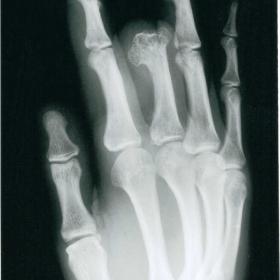
Plain radiography showed hypertrophy of soft tissues of the stump of right middle finger with flaring of distal end of proximal phalanx secondary to osteophytosis. Periarticular osteophytes were present in the middle phalanx of right index and ring finger.
MR imaging was performed on a Philips Gyroscan 0.5 T scanner. Axial T1 spin echo, coronal T1 gradient, coronal T2 spin echo, axial and coronal STIR sequences were performed using 4mm-slice thickness. Based on the MR appearances, the diagnosis of Macrodystrophia Lipomatosa (MDL) was made. The patient is awaiting surgery to reduce the bulk of the affected digit to improve cosmetic and function of the hand.
MDL was first described by Feriz in 1925, who referred only to localised gigantism in the lower extremity. In 1960 Golding suggested that the term was also applicable to similar involvement of the upper extremity. MDL shows no sex predilection, even though the earlier reports suggested that it is more often seen in female’s (1).
MDL is a rare form of localised gigantism noted soon after birth and affects one or more digits, usually in a single extremity; however, an entire extremity may be affected. The lower extremity is more commonly involved than the upper extremity. Most cases involve the middle and index finger, corresponding to the territory supplied by the branches of the median nerve (2). Digital overgrowth ceases at puberty and the usual reason for surgical correction is cosmetic appearance, although mechanical problems can be encountered when secondary degenerative joint disease reduces function and large osteophytes result in compression of the neurovascular structures. The aetiology remains obscure and there have been many proposed theories including degeneration and autonomic dysfunction(3).
The pathological findings in MDL are predominantly infiltration and hypertrophy of adipose tissue within subcutaneous tissue, nerve sheaths and periosteum. Periosteal involvement leads to the localised overgrowth of bone. Phalangeal enlargement is caused by endosteal and periosteal deposition of bone. Neural enlargement is due to infiltration of fibroadipose tissue into the nerve sheath itself and not by an increase in the size or number of axons(3).
Plain radiography shows diffuse overgrowth of soft tissues ventral to the enlarged digits, which commonly extends lateral to the involved bone in the hand and medially in the foot. The fat deposition is most marked in the distal end of a digit and may extend into the palm of the hand or sole of the foot, in which case the adjacent bones may be enlarged. Both the length and width of the bones are increased. The phalanges are usually curved medially in the hand and laterally in the foot. Flexion may be impaired and the phalanges held in hyperextension. Soft tissue lucencies may be seen which reflect the dramatic overgrowth of fat characteristic of this lesion(1).
On MRI, the lesion demonstrates high signal intensity on T1 weighted image and intermediate on T2 weighted image, similar to normal fat, admixed with areas of low signal due to fibrous hypertrophy(4). These findings in the presence of macrodactyly are considered diagnostic of MDL.
The majority of the differential diagnosis for macrodactyly such as Neurofibromatosis, lymphangiomatosis, hemangiomatosis and Klippel-Trenaunay-Weber syndrome can be differentiated on clinical grounds. On MR the differential diagnosis is limited to Fibrolipomatous hamartoma of the nerve (FLH) and proteus syndrome. Fibrolipomatous hamartoma can be seen in association with macrodactyly and displays fat deposition within the nerve sheath(5), whereas MDL demonstrates fat deposition within the nerve sheath, bone marrow, periosteum, muscles and subcutaneous tissues. The relationship between FLH and MDL may be confusing. Fibrolipomatous hamartoma of the nerve is one of the features of MDL and approximately 66% of cases of FLH are associated with macrodactyly (5). FLH may occur without macrodactyly. MDL does not share the cerebroid thickening of palms and soles or the lineal skin lesions characteristic of proteus syndrome.
In summary, MR appearance of macrodystrophia lipomatosa is characteristic and no further investigation is necessary for the diagnosis.
Macrodystrophia Lipomatosa


Based on the provided X-ray and MRI images, marked thickening and enlargement of the distal and partially proximal soft tissue and bone are observed in the right middle finger, with specific features as follows:
Considering the progressive thickening and deformation of the affected finger, together with clear adipose tissue hyperplasia on imaging, excessive bone growth, and nerve sheath involvement, possible diagnoses include:
Based on the patient’s background—32-year-old male, continued enlargement of the right middle finger despite partial amputation, with MRI findings predominantly demonstrating abnormal adipose overgrowth involving the nerve sheath and excessive bone growth—these match the literature descriptions of Macrodystrophia Lipomatosa (MDL).
Hence, the most likely diagnosis is: Macrodystrophia Lipomatosa (MDL).
FITT-VP Principle Reference:
Adjust the frequency (Frequency), intensity (Intensity), time (Time), type (Type), progression (Progression), and volume (Volume) of training according to functional recovery. Gradually increase exercise load under the guidance of rehabilitation therapists or hand surgeons to ensure safety.
Disclaimer: This report is a reference analysis based on existing imaging and medical history and cannot replace in-person consultation or professional medical advice. Specific treatment plans should be made in consideration of the patient’s actual condition under the guidance of qualified physicians.
Macrodystrophia Lipomatosa