Chondrosarcoma of the Scapula


Clinical History
The patient presented with aching pain in the left shoulder. Shoulder radiography was obtained, which revealed an abnormality prompting further evaluation with skeletal scintigraphy and MR imaging.
Imaging Findings
The patient presented with aching pain in the left shoulder. Shoulder radiography was obtained, which revealed an abnormality prompting further evaluation with skeletal scintigraphy and MR imaging.
Radiography (Fig. 1A) revealed an osteolytic lesion with popcorn calcifications and a sclerotic margin, measuring 3.5 x 4.1 cm, and involving the mid to inferior scapular body. Skeletal scintigraphy (Fig. 1B) showed focal increased uptake at the medial scapular border. At MR imaging, the lesion demonstrated ring and arc contrast enhancement, cortical destruction, and demonstrated signal characteristics consistent with chondroid matrix (Fig. 1C).
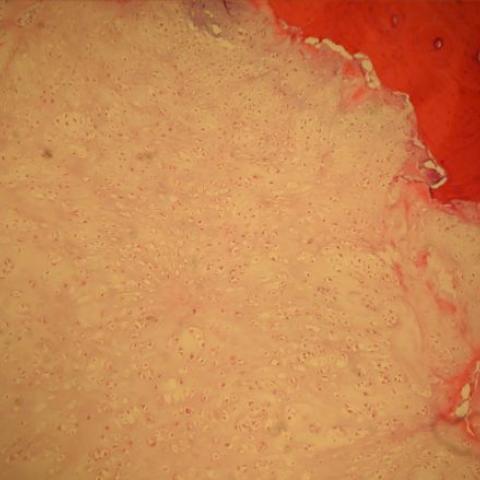
The patient underwent partial scapulectomy for tumor removal. Grossly, the mass involved cortical bone and appeared to extend into the adjacent soft tissues. Near the center of the mass there was a dense, irregular calcification. Histopathologic examination (Fig. 1D) showed a cartilaginous neoplasm with lobular architecture, well-preserved chondroid matrix, and variable cellularity. Most of the cells appeared uniform with hyperchromatic nuclei and absence of significant nuclear pleomorphism or mitotic figures. A permeative growth pattern with focal cortical bone penetration was also noted.
Discussion
Chondrosarcomas are malignant neoplasms of cartilage-forming cells and are also the third most common primary malignant tumor of bone. They are the most common malignant neoplasm of the scapula and may arise as a primary bone tumor or as a secondary lesion from a pre-existing enchondroma or exostosis. Most patients with scapular chondrosarcoma present with shoulder pain. Chondrosarcomas can also arise secondarily from other preexisting lesions to include enchondromas, osteochondromas, Paget’s disease, and fibrous dysplasia. The different types of primary chondrosarcoma include conventional intramedullary, clear cell, juxtacortical, myxoid, mesenchymal, extraskeletal, and dedifferentiated. The conventional type is the most common, involving the diaphysis or pelvis in the majority of cases (1).
Radiography usually demonstrates a lytic and sometimes expansile process, and can resemble an aneurysmal bone cyst or giant cell tumor (true aneurysmal bone cysts typically occur in a younger age group and do not contain popcorn calcifications). Periosteal reaction is often seen with chondrosarcomas and is an indication of a more aggressive process. Chondrosarcomas typically will contain ring and arc or popcorn-like calcifications, which can be seen radiographically and at cross-sectional imaging. In general, it is the high grade lesions that result in cortical bone destruction, deep endosteal scalloping, and soft tissue extension. Skeletal scintigraphy usually will show increased uptake. On MR imaging, calcifications appear hypointense on all pulse sequences, while cartilaginous matrix manifests as high T2 and low T1 signal areas, representing the high water content of the hyaline cartilage (2). High grade chondrosarcomas tend to show more variable MR appearance. The typical lobular appearance of chondroid matrix is not seen with the dedifferentiate type. Low-grade chondrosarcomas have been notoriously difficult to differentiate from enchondromas. However, a recent study by Geirnaerdt et al concluded that the combination of early and exponential enhancement correlated significantly with a malignant cartilage-forming tumor (3).
Histologically, chondrosarcoma cells are binucleated with myxoid areas. Permeative borders and invasion of soft tissue are characteristic features and can help to distinguish chondrosarcomas from enchondromas (4).
Local staging is needed to display the extent of the tumor in relation to anatomical landmarks for surgical planning. The standard treatment is surgical excision with the goal of leaving a wide margin; limb sparing resection is associated with a good prognosis for grade I chondrosarcomas. When a grade I chondrosarcoma recurs, it is usually considered then to be a high-grade lesion (5).
Differential Diagnosis List
Final Diagnosis
Low-grade chondrosarcoma of the scapula
Liscense
Figures
Conventional Radiography of the Scapula

Skeletal Scintigraphy

MRI Scapula

Histology
Medical Analysis Report
I. Imaging Findings
1. X-ray (Left Shoulder): A relatively well-defined mass within the scapula shows localized erosive changes. “Ring-shaped” or “popcorn-like” calcifications suggest a calcified cartilaginous matrix.
2. Bone Scan: The corresponding area of the left shoulder reveals increased radionuclide uptake, indicating an active metabolic lesion or neoplastic process.
3. Magnetic Resonance Imaging (MRI): The lesion appears markedly hyperintense on T2-weighted images and iso- to hypointense on T1-weighted images. Areas of cartilaginous matrix are visible, and the calcified portions demonstrate reduced signal across sequences. There is some cortical destruction and mild soft tissue involvement. Overall, these imaging findings suggest a chondroid tumor with invasive features.
II. Potential Diagnoses
-
Chondrosarcoma:
Given its typical occurrence in the scapula region, the “ring” or “popcorn-like” calcifications on imaging, tumor expansion, and cortical destruction, chondrosarcoma is the primary consideration. -
Enchondroma:
Also presents with an intramedullary cartilaginous lesion and calcifications but usually shows lower biological activity, less local invasion, and minimal cortical destruction or soft tissue mass. -
Aneurysmal Bone Cyst (ABC):
Often a cystic, expansile lesion found in younger individuals. It does not typically feature “chondroid” calcifications. Some imaging overlap is possible, yet the specific calcification pattern seen here is not typical for ABC. -
Giant Cell Tumor (GCT):
Usually occurs in skeletally mature young adults, often around the epiphyseal-metaphyseal region of long bones. It lacks prominent cartilaginous calcification and is most commonly located in the ends of long bones, which is less consistent with this presentation.
III. Final Diagnosis
Based on the patient's age (19 years), the location in the scapula, clinical symptoms (left shoulder pain), imaging findings (typical cartilaginous calcification and invasive changes), and histopathological features (e.g., binucleated chondrocytes, chondroid matrix with myxoid changes), the most likely diagnosis is: Scapular Chondrosarcoma.
IV. Treatment Plan and Rehabilitation
1. Treatment Strategy:
(1) Surgical Treatment: The standard approach involves wide or marginal extended resection, aiming to preserve as much shoulder and scapular function as possible. If necessary, partial resection of the scapula with reconstruction may be performed.
(2) Adjuvant Therapy: For higher-grade or more locally invasive tumors, postoperative radiotherapy or chemotherapy can be considered. However, for most low-grade chondrosarcomas, surgery is the mainstay of treatment and radiotherapy or chemotherapy has limited benefit.
(3) Follow-up: Routine imaging follow-up to monitor for local recurrence or distant metastases is crucial, especially in the first 5 years.
2. Rehabilitation/Exercise Prescription Recommendations:
After surgery, and based on the extent of resection and muscle strength, gradually progress with shoulder joint and upper limb functional training. Pay special attention to any reconstruction or support devices in place to maintain scapular or shoulder stability. Follow the FITT-VP principle:
- Frequency: In the early postoperative phase, perform joint mobility exercises and light muscle-strengthening 1–2 times daily. After 3–4 weeks, transition to 3–5 sessions per week.
- Intensity: Begin with low-resistance, low-load exercises to avoid undue tension and instability. Gradually increase resistance according to pain tolerance and muscle strength.
- Time: Start with short sessions (10–15 minutes) and progressively increase to 30 minutes or more per session, dividing the exercises into sets. Avoid continuous, lengthy sessions initially.
- Type: Progress from passive range-of-motion to active exercises, incorporating resistance bands and lightweight dumbbells. Include isometric and isotonic exercises targeting the muscles around the shoulder joint.
- Progression: As range of motion and muscle endurance improve, gradually increase the range and weight load. Later stages may include low-impact activities like swimming or use of a rowing machine to safeguard shoulder stability.
- Volume: Carefully increase the number of sets and repetitions as tolerated, ensuring that training overload does not cause excessive fatigue or risk re-injury.
Throughout rehabilitation, closely monitor wound healing, pain levels, and changes in joint mobility. If significant pain, swelling, or functional deterioration arises, promptly seek medical evaluation.
Disclaimer: This report is based solely on the available imaging and initial pathology information and offers reference analysis only. It does not replace an in-person consultation or professional medical advice. Specific treatment and rehabilitation should be determined by the attending physician in accordance with the patient’s actual condition.
Human Doctor Final Diagnosis
Low-grade chondrosarcoma of the scapula