

An 87 year old lady presenting with isolated ipsilateral greater and lesser trochanter fractures following a simple fall.
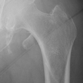
An 87 year old lady presented to the Accident and Emergency department following a syncopal event and pain in the left hip. Whilst at home, she stood up in her bedroom, felt dizzy and passed out. On waking up on the floor, she found that she could not weight bear due to pain in the left hip. She has a past medical history of osteoporosis and asthma but no cardiac history. Prior to falling she was living alone and mobilised without any walking aids. On examination there was no limb length shortening or external rotation. There was pain on palpation over the greater trochanter. Internal and external rotation was painless but flexion to 45 degrees caused discomfort. AP and lateral radiographs of the left hip revealed fractures through the greater and lesser trochanters of the femur but no fracture through the femoral neck (figures 1 and 2). The above findings were also confirmed with CT scans (figures 3 and 4). The decision was made to treat this conservatively because the absence of a fracture through the femoral neck did not compromise the load bearing capacity of the femur. Once mobile, she was discharged home without further complication.
Greater and lesser trochanter fractures usually occur in isolation. With regards to greater trochanter fractures, there are two distinct types occurring in different age groups. Firstly, there are epiphyseal separations seen in the 7-17 years age group and the second is a comminuted fracture seen in adults and usually only part of the trochanter is generally involved(1). The latter is usually due to a direct blow to the greater trochanter. Isolated avulsion fractures of the lesser trochanter occur below the age of 20 in 85% of cases (2). The injury is usually an apophyseal avulsion and is secondary to a forceful contraction of the iliopsoas muscle. It is also infrequently seen in elderly patients with osteoporosis and is believed to be due to rarefecation of the trabecular structure of the lesser trochanter leading to loss of resistance of iliopsoas contraction (2). Non traumatic avulsion fractures to the lesser trochanter can also occur with metastatic bone disease (3). Ipsilateral lesser and greater trochanter fractures are very rare and there is only one reported case in the medical literature (4). Interestingly, the events surrounding the cause of the fracture were similar to those in our case in that a 91 year old gentleman had fallen, had no recollection of events and was found on the floor by his carers at home. Unfortunately, this meant that the mechanism of injury was uncertain also. In our case, the absence of an intertrochanteric extension allowed the patient to weightbear as there was no discontinuity between the femoral head and the shaft. MRI has been proven to be a more sensitive tool in diagnosing occult hip fractures than other modalities of imaging including CT and isotope bone scan (5, 6). However in the case we describe, we are confident that there was no additional intertrochanteric extension as such a fracture pattern would have been extremely unstable and displacement would have been inevitable on weight bearing.
Greater and lesser trochanter fractures of the hip.


Based on the provided X-ray and CT images, a clear fracture line can be observed in the proximal femur (both the greater trochanter and lesser trochanter areas), and both fractures are on the same side (same-side fractures of the greater and lesser trochanters). On the X-ray, the fracture lines are confined to the regions of the greater and lesser trochanters, without any obvious involvement of the intertrochanteric area or disruption between the femoral neck and femoral shaft. CT reconstruction further confirms the separation at both fracture sites, yet no significant displacement or severely separated fragments are noted. Additionally, no other bony structures show evident abnormal signals.
Furthermore, the images suggest reduced bone density, indicating osteopenia or osteoporosis. There is no obvious large-scale soft tissue swelling or significant hematoma, nor are there imaging characteristics suggestive of secondary lesions (such as metastatic tumors).
Taking into account the patient’s advanced age, history of a fall, clinical symptoms, and imaging evidence, the most likely diagnosis is: “Isolated Fractures of the Greater and Lesser Trochanters on the Same (Right/Left) Side”. Imaging does not indicate that the fracture extends clearly into the intertrochanteric region, nor are there signs of pathological fractures. This is likely the result of a simple trauma on a background of osteoporosis.
After confirming the stability of the fracture or completing appropriate treatment, a gradual rehabilitation plan should be developed:
Disclaimer: This report is a reference analysis based on available information and cannot replace an in-person consultation or professional medical advice. The patient’s specific diagnosis and rehabilitation plan must be determined by a clinical physician according to individual circumstances.
Greater and lesser trochanter fractures of the hip.