Multicentric hemangioendothelioma involving multiple bones of the foot


Clinical History
Swelling and pain of the left extremity .
Imaging Findings
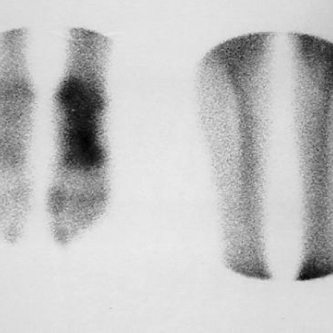
A patient 35 years old was hospitalized with swelling and pain of the left foot . His past medical history was free for any injury or accident . CT of the extremities was revealed osteolytic regions ,geographic type - IB without periosteal reaction , of the bones of the left foot . These regions were noted in the calcaneum , cuboid and the base of the 5th metatarsal bone (Fig 1a,b). The MRI findings were similar (Fig 2a,b,c). The bone scan revealed uptake of the bones of the left foot and knee (Fig 3a,b). The treatment was surgical and the histological findings confirmed the diagnosis of multicentric hemangioendothelioma ( Fig 4a,b,c,d).
Discussion
Hemangioendothelioma is a rare malignant tumor of bone . It is composed of irregular anastomosing vascular channels lined by one or several layers of atypical endothelial cells (1) . It is the endothelial nature of the proliferating cell that distinguishes this lession from a hemangiopericytoma , in which pericytes represent the cell of origin . Hemangioendotheliomas (angiosarcomas) are more frequent in men than in women , in a ratio of approximately 2 to 1 (1,2,3,4) .The lesions are observed in patients of all ages , althouh the majority of affected persons are in the 3rd , 4th or 5th decade of life . Those with multifocal disease usually are about 10 years younger than those with single lesions (5). Local pain and swelling are the two most characteristic clinical findings and may be of weeks' , months' or even years' duration . Hemangioendothelioma predominates in the long tubular bones , especially those in the lower extremity (6,7,8,9,10).The long bones are affected in approximately 60% of cases , with preferential involvement of the tibia 23%, femur 18% and humerus 13% , pelvis 7% , skull 4% . The ribs are affected in approximately 5% of cases (11,12,13,14,15) . Rare sites of involvement are the scapula , clavicle , sternum , radius , patella and the small bones of the hand and foot (16,17) . A metaphyseal or diaphyseal location is typical . One of the characteristic features of hemangioendothelioma is synchronous or metachronous multicentric disease ( 20 - 50% of cases ) .Multiple lesions may occur in a single bone or one or more tumor foci may be apparent in multiple bones in a single extremity - especially the lower extremity ). Angiosarcomas may develop in bones with preexisting abnormalities , such as chronic osteomyelitis , osteonecrosis , neoplasms or at sites of metallic fixation devices (18,19,20) . The principal radiographic pattern is osteolysis that uncommonly is accompanied by osteosclerosis .The lesions are variable in size . Multifocal involvement is an important roentgenographic pattern of this neoplasm . It may be manifested as two or more osteolytic lesions involving a long segment of a single bone or osteolysis in contiguous bones . The radiographic demonstration of multiple neoplastic foci in the cortical bone or spongiosa , or both , leading to a bubble -like appearance and osseous expansion without periostitis in a tubular bone of a lower extremity is highly characteristic . Angiosarcomas may lead to extensive destruction of several carpal or tarsal bones or rarely phalanges or metacarpal and metatarsal bones . Other diagnostic considerations include skeletal metastasis, plasma cell myeloma , cystic angiomatosis , histiocytosis , Kaposi's sarcoma , bacillary angiomatosis and fungal or tuberculous osteomyelitis .Bone scintigraphy also may be useful in confirming involvement of the bones of a single extremity in patients with angiosarcomas . Treatment involves surgical ablation . Some patients have responded favorably to radiation therapy .When a patient is diagnosed as having hemangioendothelioma of bone , a search should be made for multicentricity .
Differential Diagnosis List
Final Diagnosis
Multicentric Hemangioendothelioma involving multiple bones of the left foot .
Liscense
Figures
Computed tomography . Osteolytic lesions in small bones of the foot .


MRI


Histopathologic findings of hemangioendothelioma


scanning of the bones

Medical Analysis Report
I. Imaging Findings
From the provided CT, MRI, and radionuclide bone scan images, there are localized multiple osteolytic lesions in the left foot (including the ankle joint and tarsal regions), with irregular margins showing foam-like or cystic translucent areas. The cortical bone may appear focally thinned or mildly expanded. No obvious periosteal reaction or typical “onion-skin”/“sunburst” patterns are noted. On MRI, the lesion exhibits mixed signal characteristics, predominantly high signal on T2-weighted images, suggesting possible vascular or other soft tissue components.
When lesions primarily involve long bones or tarsal bones, one should be alert to the possibility of multiple foci. The patient’s left foot soft tissue shows mild swelling or signs of passive displacement, but no large intra-soft-tissue mass is evident. The radionuclide bone scan indicates increased radiotracer uptake in the same region of the left foot, hinting at high local metabolic activity and raising concern for malignancy.
II. Potential Diagnoses
Considering the patient’s age (35 years old, male), clinical symptoms (local swelling and pain), osteolytic changes on imaging, and pathological findings, the following differential diagnoses should be considered:
- 1. Bony Hemangioendothelioma (Angiosarcoma): This disease typically shows multiple “foam-like” osteolytic lesions on imaging, often seen in middle-aged individuals, and is confirmed by positive vascular markers such as CD31.
- 2. Bone Metastases: Many solid tumors can present with osteolytic metastases; however, they are usually accompanied by a known primary tumor history, and immunohistochemical studies typically do not reveal vascular endothelial markers.
- 3. Multiple Myeloma: Commonly seen as multiple osteolytic lesions among older patients, frequently associated with abnormal blood counts and immunoglobulin levels, which does not match the vascular endothelial marker positivity in this case.
- 4. Other Vascular or Infectious Lesions: For instance, cystic hemangioma, fungal or tuberculous osteomyelitis. These may also demonstrate bony destruction and osteolytic lesions but can usually be distinguished via clinical and pathological evaluations.
III. Final Diagnosis
Integrating the patient’s history, imaging findings of multiple osteolytic lesions, and histopathological evidence demonstrating irregular vascular channels with endothelial cell proliferation (CD31 and reticulin staining support), the final diagnosis is highly consistent with angiosarcoma (bony hemangioendothelioma). This disease often presents with multifocal involvement, aggressiveness, and a high tendency for recurrence or metastasis. Further evaluation is needed to confirm the absence or presence of additional lesions in other bones or soft tissues.
IV. Treatment Plan and Rehabilitation Program
1. Treatment Strategy:
- Surgical resection or curettage of the lesion is the primary treatment. If there is significant local bone destruction, bone grafting or internal fixation may be considered. For extensive or multifocal lesions, combined radiotherapy can be recommended.
- In certain advanced or metastatic cases, chemotherapy may be considered. However, the overall response to chemotherapy is limited; decisions should be based on pathological staging and individual circumstances.
- Adjuvant radiotherapy before or after surgery can help reduce the risk of local recurrence.
2. Rehabilitation and Exercise Prescription:
-
Early Stage (Postoperative Recuperation):
- Focus on protective immobilization and elevation of the affected limb to reduce edema.
- Begin passive range-of-motion exercises to prevent soft tissue contractures, along with gentle, non-weight-bearing movements such as ankle and toe flexion-extension.
-
Intermediate Stage (Gradual Weight-Bearing):
- As bone healing and soft tissue recovery permit, gradually progress to partial weight-bearing walking or using assistive devices, according to medical advice.
- Perform active joint movements combined with light resistance training (e.g., resistance bands, light dumbbells) to strengthen the lower limbs.
-
Late Stage (Functional Recovery and Coordination Training):
- Gradually resume normal weight-bearing and gait training. Depending on the stability of the lesion, low-impact walking or slow jogging, single-leg balance exercises, and muscle strengthening can be introduced.
- Subject to the patient’s cardiopulmonary function and bone strength, add cycling or swimming to improve overall cardiovascular endurance.
-
FITT-VP Principles:
- Frequency: 3–5 times per week.
- Intensity: Low intensity initially, with a subjective effort not exceeding 3–4 on a scale (mild to moderate). Progress to moderate intensity as recovery advances.
- Time: Start with 10–15 minutes per session and gradually increase to 30–45 minutes.
- Type: Focus on exercises that protect the affected limb, including range-of-motion, low-impact aerobic activities, and limited resistance training.
- Progression: Increase load and duration by stages according to bone and soft tissue healing, closely monitoring for pain, swelling, or other unusual symptoms.
-
Important Considerations:
- Avoid vigorous or high-impact activities during healing or rehabilitation to prevent fractures, displacement, or risk of tumor recurrence.
- If significant pain, local redness, or a sudden decrease in range of motion occurs, seek medical evaluation promptly.
Disclaimer:
This report is based on the available medical records and imaging data for reference purposes and cannot replace in-person consultations or professional medical advice. If you have any questions or develop new symptoms, please consult a specialist without delay.
Human Doctor Final Diagnosis
Multicentric Hemangioendothelioma involving multiple bones of the left foot .