Tuberous sclerosis of the tibia


Clinical History
A 12 year old girl with known tuberous sclerosis presented with a valgus deformity of the left tibia.
Imaging Findings
A 12 year old girl with known tuberous sclerosis presented with a few months history of a worsening valgus deformity of the left lower leg. She had had previous neurosurgical procedures including removal of a giant cell astrocytoma from the brain, and insertion of a VP shunt to relieve bilateral hydrocephalus. She had cognitive impairment and was on medication for epileptic seizures. Plain radiographs were performed of the affected limb. They confirmed the valgus deformity and showed periosteal thickening of the left tibia. The left fibula was also affected to a lesser extent.
Discussion
Tuberous sclerosis is an autosomal dominant condition classically characterized by a clinical triad of epileptic seizures, mental retardation and adenoma sebaceum. This triad is found in only 30% of patients and the disorder has a wide spectrum of phenotypic expressions with hamartomatous malformations occurring mainly in the brain, kidney, lung, skin and heart. The extracranial skeleton may be focally or diffusely involved with radiolucent cyst-like lesions or dense sclerotic deposits. Irregular subperiosteal new bone formation can produce a thickened undulating cortical contour. Cortical involvement is usually seen in the short tubular bones of the hands and feet, and occasionally, the long tubular bones. Radiolucent cyst-like lesions mainly occur in the distal phalanges of the hand. Significant deformity is treated with orthopaedic procedures, and a tibial osteotomy will be performed on this individual. The prognosis for restoration of skeletal function is good. The differential diagnosis of the plain radiographs is tuberous sclerosis or neurofibromatosis. Previous infection or trauma is less likely.
Differential Diagnosis List
Final Diagnosis
Tuberous sclerosis of the tibia (and fibula).
Liscense
Figures
AP radiograph of the left tibia and fibula.
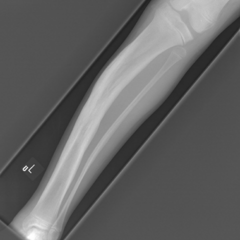
Lateral radiograph of the left tibia and fibula.

1. Radiological Findings
Based on the provided left lower leg anteroposterior and lateral X-ray images, the following main features are observed:
• The left tibial shaft shows a mild to moderate valgus deformity (the mechanical axis of the tibia deviates laterally), consistent with the clinically reported tibial valgus deformity.
• Overall bone density does not appear markedly reduced, but locally the cortical bone shows irregular morphology, slightly thickened or undulating margins.
• No obvious acute fracture line or displaced fracture fragments are seen; no significant soft tissue swelling or periosteal reaction suggesting acute inflammation.
• In conjunction with the patient’s history of tuberous sclerosis, note that long bones can present changes related to tuberous sclerosis, such as local cortical thickening and irregular cortical contours.
2. Potential Diagnoses
Based on imaging features and the patient’s underlying condition, the primary considerations include:
• Tuberous Sclerosis:
Correlates with the patient’s known history; can present localized cortical changes, undulating thickening, and bone deformities, including valgus deformity.
• Neurofibromatosis:
Another hereditary disorder that can lead to skeletal deformities and cortical changes in long bones. Some patients may present with similar long bone angular deformities. Differentiation is required.
(As the patient’s history has already confirmed tuberous sclerosis, other possibilities such as post-infection changes or post-traumatic deformities are less consistent with the clinical and radiological features and can be excluded.)
3. Final Diagnosis
Taking into account the patient’s age, established diagnosis of tuberous sclerosis, and imaging findings of a tibial valgus deformity with mild irregular cortical thickening, the most likely diagnosis is:
Tibial valgus deformity due to tuberous sclerosis.
If further clarification of the local bony involvement is required, additional imaging studies such as CT or MRI can be considered to evaluate soft tissue, cortical, and medullary changes; bone biopsy might be considered in certain cases, though it may not be necessary given the clinical history.
4. Treatment Plan and Rehabilitation
- Surgical Treatment: Due to the significant tibial valgus deformity and potential malalignment, tibial osteotomy (closed or open wedge approach depending on the degree of deformity) with internal fixation is recommended to correct the mechanical axis of the tibia.
- Postoperative Rehabilitation and Exercise Prescription:
- Early Stage (0–6 weeks postoperative): The main focus is on surgical wound healing and pain management. Perform basic lower limb muscle maintenance exercises, avoiding high-impact weight-bearing. Under medical guidance, engage in isometric exercises (e.g., quadriceps sets) and simple ankle joint range of motion exercises.
- Intermediate Stage (6–12 weeks postoperative): If bone healing is satisfactory, gradually introduce weight-bearing exercises, such as partial weight-bearing ambulation with crutches. Begin low-intensity lower limb strengthening and stability exercises (e.g., straight leg raises, suspended leg exercises) two to three times a week, without causing significant pain or swelling.
- Late Stage (3–6 months postoperative): Transition to strengthening and functional restoration, progressively increasing resistance for lower limb strengthening, balance training, and light aerobic activities (e.g., slow cycling, brisk walking). Frequency of three to four times a week, with exercise duration gradually increased from 15–20 minutes to over 30 minutes, according to the patient’s tolerance.
- Long-term Stage (beyond 6 months postoperative): Depending on bone healing and functional recovery, most daily activities and moderate exercise can be resumed. Higher-intensity activities, such as running, may be reintroduced with close monitoring of the lower limb alignment and ankle stability.
- Application of the FITT-VP Principle:
- Frequency: Starting with two to three sessions per week early on, progressing to three to four sessions per week in the intermediate phase.
- Intensity: Gradually increase based on pain and fatigue levels, ensuring no significant discomfort or excessive stress on bone structures.
- Time: Increase session duration from 10–15 minutes up to 30 minutes or longer.
- Type: Combination of lower limb strengthening, joint mobility, and aerobic conditioning. Begin with non-weight-bearing or partial weight-bearing exercises and progress to full weight-bearing over time.
- Volume: Increase overall workload in accordance with recovery progress, monitoring fatigue and swelling in the postoperative limb.
- Progression: As bone healing and function improve, gradually intensify and challenge the exercises to maintain and enhance muscle strength, flexibility, and weight-bearing capacity.
During rehabilitation, special attention should be paid to:
• Possible involvement of other organ systems by tuberous sclerosis, such as cardiopulmonary impairments, which may necessitate adjusting exercise intensity.
• The risk of fragile bone or delayed union; if abnormal pain or discomfort occurs, seek medical attention promptly.
• Regular postoperative follow-up (X-ray or CT) to monitor bone healing and the effectiveness of correction, allowing for individualized adjustments to the rehabilitation plan.
Disclaimer: This report is based solely on the current medical history and imaging data provided for reference by a professional medical practitioner. It should not replace a face-to-face consultation and diagnosis. The specific treatment plan and rehabilitation protocol must be determined by the attending physician based on the patient’s actual condition.
Human Doctor Final Diagnosis
Tuberous sclerosis of the tibia (and fibula).