Radiation necrosis of the humeral head: A case report.


Clinical History
The patient suffered increasing pain in the right shoulder, the clinical examination revealed a severe restriction of right shoulder movements.
Imaging Findings
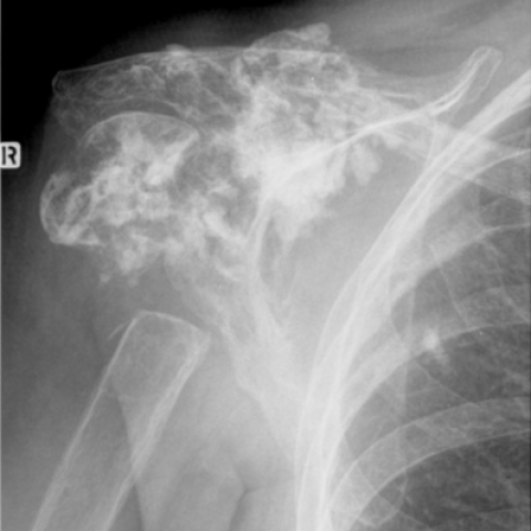
A 77 year old female suffered pain in the right shoulder. Clinical examination revealed a restriction of shoulder movements.18 Years ago, a plain radiograph visualised a lesion in the humerus that was considered as a metastasis of a previously endured breast carcinoma. Radiotherapy was started. A recent plain film demonstrated a deformation of the right shoulder with an irregular shape of the humeral head and a sharply defined pseudarthrosis at the border with the diaphysis. The remaining parts of the humeral head consisted of irregularly calcified areas, non-congruent with the articular surface. There was partial osteolysis of the acromial extremity of the clavicle, the glenoid cavity, the acromion and the lateral part of the scapula. Magnetic resonance imaging of the right shoulder demonstrated a fragmented aspect of the humeral head with soft tissue calcifications and/or fibrosis that extended twelve centimetres craniocaudally from the level of the acromioclavicular joint distally. No abnormal soft tissue mass and no infiltration of the bone marrow were visible. There were considerable hypotrophic changes (with fatty involution) of the right pectoralis and shoulder muscles. There was also a pseudarthrosis of the proximal humerus with a distance of two centimetres between both bony parts of the pseudarthrosis. Debris and granulation tissue were present in that cavity. The acromial extremity of the clavicle also displayed pseudarthrosis. Soft tissue calcifications and/or fibrosis were present in that cavity. Moreover there was a partial ankylosis of the glenoid cavity with some remnants of the right humeral head.
Discussion
Sengupta and Prathap (1973) described similar abnormalities of the humerus in three cases. The cases concerned women by whom osteonecrosis occured seven to ten years after irrradiation therapy for breast carcinoma. Kollar et al. (1967) described a case of erosion of the neck of the humerus and osteonecrosis of the scapula five years after irradiation therapy in a patient with breast carcinoma. Osteonecrosis presents years after irradiation therapy as mixed sclerotic and lytic lesions altough these lesions are initially mainly lytical (Burgener and Kormano, 1991). It concerns radiation damage to the bone and connective tissue that results in hypoxic, hypovascular and hypocellular tissue due to an increasing cell death and collagen lysis. The resulting tissue damage depends on the dosage (Howland et al., 1975; Burgener and Kormano, 1991), quality of the x-ray beam, specific bone or bones involved, length of time after therapy and the superimposition of trauma or infection (Resnick, 2002). The changes in bone after irradiation therapy can in brief be defined as atrophic (Howland et al., 1975). These changes are asymptomatic. Fractures are common complications in this atrophic bone. Three patients in the study of Howland et al. (1975) developed, without any significant recognizable trauma, a fracture of the proximal humerus after irradiation therapy, as was the case in our patient. In one patient the fracture did not heal. Such fractures frequently are associated with pseudarthrosis, considering they manifest with relatively few complaints and as such the bone fragments are insufficiently immobilized (Resnick, 2002). Non union and atrophic changes were also present in our patient. In summary we described a case of radiation necrosis, radiologically resembling osteosarcoma, but proven to be benign osteonecrosis with MRI. The atrophic changes in the bone following irradiation therapy have resulted in an insufficiency fracture (spontaneously or not ) of the proximal humerus and a fragmendted appearance of the humeral head in our patient. As a consequence of insufficient immobilization of the bone fragments a pseudarthrosis developed. Also partial ankylosis of the glenoid cavity with the remnants of the right humeral head developed. An untreated posterior shoulder luxation and an immobilization of the glenohumeral joint were responsible for the latter.
Differential Diagnosis List
Final Diagnosis
Radiation necrosis of the humeral head.
Liscense
Figures
Fig. 2

Fig. 3

Fig. 4

Fif. 1
1. Imaging Findings
This case involves a 77-year-old female patient who reports progressively worsening right shoulder pain. Clinical examination indicates a severely restricted range of motion in the right shoulder joint. Based on the provided MRI and X-ray images, the following key features can be observed:
- Significant bony destruction in the proximal right humerus, exhibiting both osteopenia and sclerosis (mixed bone changes), with an irregular local contour.
- The humeral head shows comminuted changes, irregular margins, and visible fracture edges, suggesting an old or nonunion fracture.
- Disruption of shoulder joint structure, with abnormal articulation in parts of the glenohumeral joint, indicating possible dislocation (possibly posterior) and/or malunion.
- No obvious large soft tissue mass is observed within the soft tissue region; however, chronic inflammation or fibrosis may exist around the joint, and the local soft tissue space is narrowed.
- MRI signals show localized necrotic/ischemic changes within the bone marrow (likely presenting as low or mixed signals on T1- and T2-weighted images), along with areas indicating sclerosis, consistent with radiation-induced osteonecrosis.
- Osteophyte formation and joint space narrowing are observed around the joint, suggesting degenerative changes and potential partial joint ankylosis.
2. Potential Diagnoses
Based on the imaging findings and the patient’s medical history (possibly including a history of radiotherapy post-breast cancer), the following diagnoses or differential diagnoses are considered:
- Radiation-induced Osteonecrosis
- Long-term radiotherapy history, with a combination of atrophy, sclerosis, and lytic changes in the bone.
- Typical manifestations include reduced bone vascularity, structural destruction, susceptibility to fracture, and difficulty in healing.
- Tumor or Tumor-like Lesions (e.g., Osteosarcoma)
- Secondary tumor formation is possible after radiation, but clinical and MRI findings suggest benign necrotic changes.
- If it were a neoplastic lesion, it would typically show a more pronounced soft tissue mass or rapid bone destruction; no large soft tissue mass is evident in this case.
- Degenerative Osteoarthritis (with Secondary Changes)
- The patient’s older age and significant joint degeneration may lead to bony abnormalities, but purely degenerative changes would rarely present such marked osteonecrosis.
3. Final Diagnosis
Considering the patient’s age, right shoulder pain and dysfunction, a likely history of breast cancer radiotherapy, and imaging features indicating atrophy, mixed sclerosis and lytic changes along with an unhealed old fracture, the most probable diagnosis is:
- Radiation-induced Osteonecrosis, accompanied by chronic fracture and pseudoarthrosis
Additionally, there is evidence of joint structural disarray, partial displacement of articular surfaces, and partial ankylosis, suggesting secondary changes due to an uncorrected joint dislocation or subluxation.
4. Treatment Plan and Rehabilitation Strategy
For this case of radiation-induced osteonecrosis complicated by fracture, pseudoarthrosis, and limited joint mobility, the treatment and rehabilitation strategies include:
- Conservative Management
- Pain management: Use non-steroidal anti-inflammatory drugs (NSAIDs) or appropriate analgesics to relieve pain.
- Immobilization and bracing: Consider using a shoulder brace or specialized thoracolumbar support to help stabilize the shoulder joint and reduce motion at the pseudoarthrosis site.
- Nutritional support and bone protection: Supplement with calcium, vitamin D, and medications such as bisphosphonates to slow further bone loss.
- Surgical Intervention
- If pain is unmanageable or pseudoarthrosis and nonunion lead to severe functional impairment, surgical interventions such as arthroplasty, bone graft with internal fixation, or joint replacement may be considered.
- Due to the poor blood supply associated with radiation-induced osteonecrosis, careful evaluation of surgical feasibility and healing risks is essential.
- Rehabilitation Training and Exercise Prescription
- Early Phase (Acute/Postoperative Acute Phase)
- Focus on protective activities. Avoid large-amplitude stretching and heavy load bearing.
- Perform passive shoulder range-of-motion exercises and isometric training for surrounding muscles, 2–3 times per day, 5–10 minutes per session.
- Mid Phase (Functional Recovery Phase)
- Gradually increase active range of motion within tolerable pain limits, practicing shoulder abduction, forward flexion, and external rotation.
- Depending on pain and functional improvement, combine moderate resistance band exercises 3–5 times per week for 10–15 minutes each time, avoiding excessive strain.
- Late Phase (Strengthening/Maintenance Phase)
- Gradually add functional shoulder training, for example using light dumbbells or cane-assisted exercises to enhance muscle strength and improve joint flexibility.
- Maintain a frequency of 3–5 sessions per week, progressively increasing intensity, and place emphasis on strengthening the shoulder and back muscles.
- Throughout the rehabilitation process, closely monitor the patient’s shoulder pain, swelling, and function, adjusting the intensity and methods of training as needed.
- Early Phase (Acute/Postoperative Acute Phase)
5. Disclaimer
This report is based solely on the available imaging and the provided medical history. It serves as a reference and cannot replace an in-person consultation or professional medical advice. For a definitive diagnosis and treatment plan, the patient is advised to seek further examinations and a comprehensive evaluation under the guidance of a specialized physician.
Human Doctor Final Diagnosis
Radiation necrosis of the humeral head.