Subungual exostosis of the little toe: MRI features


Clinical History
A 15 year old boy presented with generalised swelling of right little toe with no prior history of injury.
Imaging Findings
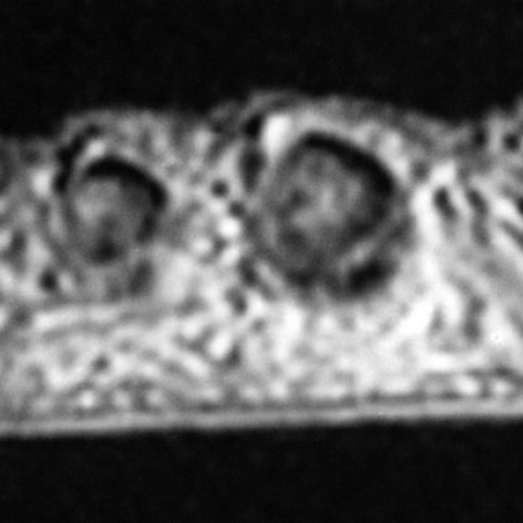
A 15 year old boy presented with a 3-month history of an enlarging, minimally painful swelling of the tip of right little toe. The swelling was generalised with no clear cut demarcations from the underlying bone and was firm in consistency with minimal tenderness on palpation. The overlying nail was thickened and spoon-shaped. No similar swellings were found elsewhere. Radiography showed bony erosion of the distal phalynx of little toe (figure 1). MRI showed a lesion enveloping the terminal phalanx of the little toe from below and laterally, with further extension of it between the dorsal surface of the bone and the nail. The lesion was low signal on the T1 sequence (figure 2a), high signal on the fat suppressed sequence (figs 2b - d) and there was no enhancement within it following gadolinium administration. No signal void to suggest calcification within the mass was demonstrated. The scan confirmed pressure erosion of the tuft of the distal phalanx. Surgical excision of the lesion was performed and histopathological assessment of the specimen showed it to be consistent with a subungual exostosis with predominant chondroid metaplasia.
Discussion
Subungual exostosis is a rare osteo-cartilaginous benign tumour arising usually from the distal phalynx of the toes, more commonly from the hallux. Very rarely they can be found in the hands. The term ‘exostosis’ is a misnomer as there is no continuity with the underlying bone. Females are affected twice as males and the lesions occur mostly during the second decade. The precise cause is not clear, but factors like trauma, chronic infection and local irritation are often implicated. The underlying pathology is the initial growth of fibro-cartilaginous tissue, which later ossifies to a varying degree. It differs from osteochondroma, which is a congenital lesion arising from the metaphysis of the underlying bone. Clinically patients may present with swelling in the distal phalynx, with some degree of tenderness often after an episode of trauma. The position of the lesion is almost always dorso-medial. Possible differential diagnoses for the lesion include osteochondroma, glomus tumour, squamous cell carcinoma and melanoma. Radiologically, the appearances can vary according to the amount of calcification. Generally the outgrowth has features of cancellous bone without a defined cortex. During the pre-ossifying stage the underlying bone can be eroded due to the compression by the tumour. Surgical excision and curettage of the base is the treatment of choice. Rarely recurrences can occur if the base is not excised completely.
Differential Diagnosis List
Final Diagnosis
Subungual exostosis of the liitle toe
Liscense
Figures
X-ray

MRI scan


Medical Analysis Report
Imaging Findings
Based on the provided imaging of the right little toe (including X-ray and MRI), the following findings are noted:
- There is an abnormal protrusion on the dorsal aspect of the distal phalanx of the right little toe, appearing as an irregular cystic or fibrous dense structure on imaging.
- On the X-ray, a mass-like shadow extends from the dorsal side of the distal phalanx, but it is not continuous with the bone shaft, and there are local signs of partial calcification or ossification.
- On MRI cross-sections, the lesion shows heterogeneous signals, possibly containing cartilaginous or fibrous components, with mild compression of the surrounding soft tissue at the lesion margins.
- There is significant swelling of the surrounding soft tissue, but no obvious fracture lines are identified, and no extensive involvement of the adjacent soft tissue is present.
Possible Diagnoses
Considering the patient’s age, clinical presentations (mild pain, no apparent history of trauma), and imaging findings, the following conditions or differential diagnoses can be considered:
- Subungual exostosis: This is common in adolescents, frequently occurring on the distal phalanx of the toes (most commonly the big toe), but may also affect other toes. The typical presentation is fibrous cartilaginous proliferation and ossification located beneath the nail bed of the distal phalanx (or under the fingernail/toenail), which is not continuous with the bone shaft.
- Osteochondroma: Usually presents as a bony protrusion outward from the bone, continuous with the metaphysis, often accompanied by a prominent bone-cartilage cap that is continuous with the bone shaft. This condition is typically congenital or developmental in origin, which does not completely match the current description.
- Glomus tumor: Often occurs under the nail bed, typically presenting with severe pain or sensitivity to local pressure. Imaging usually shows soft tissue density with marked contrast enhancement on T1/T2-weighted MRI.
- Squamous cell carcinoma and Melanoma: Both are malignant tumors that may show bone destruction or abnormal thickening of the nail bed on imaging. Clinically, they often present with bleeding, pigmentation changes, or ulceration.
Final Diagnosis
Taking into account the patient’s clinical presentation (a 15-year-old male with no significant history of trauma, mild local pain with swelling), imaging findings (dorsal proliferation of the distal phalanx, not continuous with the bone shaft, showing fibrous-cartilaginous changes), and pathophysiological characteristics, the most likely diagnosis is:
Subungual exostosis.
If there is still any doubt or further clarification is needed, surgical excision of the lesion followed by pathological examination can confirm the diagnosis and assess the nature of any tumor cells.
Treatment Plan and Rehabilitation
The most effective treatment for this condition is typically surgical excision, along with curettage of the base to reduce the risk of recurrence. The following are the recommended treatment and rehabilitation guidelines:
- Surgical Treatment and Principles
- After confirming the extent of the lesion, perform localized surgical excision to completely remove the bony outgrowth. Ensure thorough curettage of the base.
- During surgery, take care to protect the surrounding soft tissue and the nail bed structure to avoid postoperative nail bed deformities or infection.
- Postoperative Rehabilitation
To promote functional recovery of the affected toe and reduce pain and swelling, a gradual, individualized rehabilitation program should be established. The FITT-VP principle can be referenced:
- Frequency: Perform basic functional training once or twice daily.
- Intensity: Begin with mild activities, such as elevating the foot and actively flexing and extending the toes. Increase activity levels once pain subsides.
- Time: Start with 5-10 minutes of exercise each session, gradually lengthening to 15-20 minutes, depending on recovery progression.
- Type: Early stages focus on range-of-motion exercises and muscle relaxation; later stages may incorporate strengthening exercises for the lower leg muscles and foot coordination training.
- Volume: Gradually increase the frequency and duration of daily activities according to the patient’s recovery status, but avoid overexertion or aggravation of pain.
- Progression: As swelling and pain improve, introduce gentle weight-bearing activities (e.g., standing balance exercises), and consider simple gait training or other assisted exercises as appropriate.
Throughout rehabilitation, pay attention to any signs of recurrent swelling or pain in the affected toe. If significant discomfort emerges, seek timely medical consultation.
Precautions: If the patient has poor bone quality or a high sensitivity to pain, the exercise regimen should be slowed down accordingly. In cases of persistent infection or signs of poor wound healing, seek medical attention promptly for appropriate medication or other interventions.
Disclaimer
This report provides a reference analysis based on the existing medical history and imaging data, and does not replace in-person consultation or the diagnostic and treatment plans provided by a professional medical institution. If you have any questions or if symptoms worsen, please seek professional medical attention promptly.
Human Doctor Final Diagnosis
Subungual exostosis of the liitle toe