Osteoblastoma-like osteosarcoma with local recurrence in the distal radius


Clinical History
A 29-year-old female with a history of osteoblastoma of the right radius that was previously treated with an intralesional curettage was presented to the orthopaedic surgeon with recurrent pain during movement of the wrist three years after the operation.
Imaging Findings
X-Ray imaging revealed a new occurrence of osteolytic lesions around the operated area. Magnetic resonance imaging (MRI) showed a local recurrence of several newly-formed lesions in the distal meta-diaphysis, which were hypointense on T1-weighted images (T1W) and hyperintense on T2-weighted images (T2W) with centrally enhanced contrast uptake. Radiologic features of the lesions were suspected to be either a dedifferentiation from osteoblastoma to a low-grade sarcoma with skip lesions, or a local recurrence of the osteoblastoma with surrounding osteoblastomatosis (Fig. 1, 2, 3). Positron Emission Tomography and Computerised Tomography (PET/CT) was performed for staging and showed metabolic activity only in the lesion of the distal radius (SUV max. 4,2) (Fig. 4). The patient was treated with a wide surgical resection of the distal radius, a reconstruction with ulna transposition and an arthrodesis (Fig. 5). Histological results identified the lesion as a grade 1 osteoblastoma-like osteosarcoma (OBLOS).
Discussion
OBLOS is a rare variant of conventional osteosarcoma (OS) and represents 1,1 % of all osteosarcomas [1, 2]. It affects young adults with a slightly higher male-to-female ratio (1,2:1) [1, 3]. It causes chronic progressive pain in the affected area accompanied by swelling and a reduced mobility of the adjacent joint [1, 3-5]. OBLOS is hard to distinguish from a benign osteoblastoma (OB) because of their clinical resemblance and histological and radiological appearance. Unlike OB, OBLOS has the capability to metastasize and represents a high mortality risk [3]. Frequent location of OBLOS is the metaphysis of long bones, mostly the tibia, followed by the vertebrae, hands, feet and the femur [1].
Conventional radiologic imaging methods generally display the lesions as osteolytic, with thinned surrounding cortex and cortical destruction or as well-defined lesions, including a sclerotic rim [1, 3]. An MRI scan is a reliable diagnostic tool for assessing the extent of the intramedullary and soft tissue involvement. The lesions are hypointense on T1W and iso- or hyperintense on T2W, with substantial bone marrow oedema. The scans tend to show incoherent contrast enhancement centrally. However, MRI scans are inconclusive, since benign lesions may appear aggressive as well [1, 3, 6]. Histologically, OBLOS, may closely resemble osteoblastoma, sometimes with the only difference being increased mitotic activity (featuring atypical mitoses) and permeative growth in the former (OBLOS) but not the latter (osteoblastoma). On a molecular genetic level, more than 90% of osteoid osteomas and osteoblastomas but no osteosarcomas have been shown to harbour FOS or FOSB gene rearrangements [7, 8].
In this case, histopathological evaluation of the resected distal radius revealed seven separate foci of osteoblastic proliferation, morphologically resembling multiple nidi of osteoid osteoma/osteoblastoma, but focally displaying permeative growth and increased mitotic activity (Fig. 6), including some atypical mitoses. Molecular genetics was also performed and revealed no rearrangements in the FOS or FOSB genes. Hence, the histological diagnosis favoured a grade 1 osteoblastoma-like osteosarcoma (OBLOS) over osteoblastomatosis.
Prognosis of OBLOS is unfavourable because of its capability to metastasize, mostly in the lung, occasionally in the skeleton [4, 9, 10]. A wide surgical resection with chemotherapy is normally the treatment of choice [9, 11].
We report a case of OBLOS to emphasize, how difficult is its differentiation from a benign lesion. Incorrect diagnosis can lead to inadequate surgical treatment and a local recurrence. Clinicians should provide frequent follow-up visits.
Written informed patient consent for publication has been obtained.
Differential Diagnosis List
Final Diagnosis
Osteoblastoma-like osteosarcoma
Liscense
This work is licensed under a Creative Commons Attribution-NonCommercial-ShareAlike 4.0 International License.
Figures
AP radiograph at initial admission

MRI scan at initial admission


MRI scan three years after first treatment


FDG PET-CT bone scan three years after the treatment

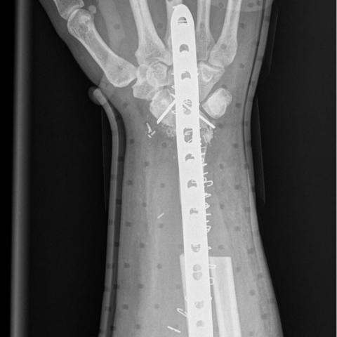
AP radiograph of the right wrist after surgical resection
Gross resection and histopathological images


Medical Analysis Report
I. Radiological Findings
Based on the provided X-ray and MRI images, multiple “osteogenic lesions” can be observed in the distal region of the right radius. These lesions have the following characteristics:
- On the X-ray images: a relatively well-demarcated area of bone destruction can be seen in the distal radius, with some areas showing thinning of the cortical bone, local sclerosis or destruction, and various degrees of osteoproliferation.
- On MRI sequences: the lesions show low or slightly low signal on T1-weighted images and iso- or slightly high signal on T2-weighted images, with localized bone marrow edema. Contrast-enhanced scans reveal uneven or scattered enhancement within the lesions, suggesting active osteogenesis or tumor cell proliferation.
- The images show multiple “nodular” or “mass-like” lesions, with some extending toward the nearby soft tissues, but primarily involving bony changes.
- Taking into account the patient’s history, postoperative changes (plate fixation and local defect repair) can be noted in the region.
Overall, the radiological findings indicate multiple areas of bone destruction with new bone formation in the distal right radius. Further correlation with clinical history and pathology is required.
II. Differential Diagnoses
Based on the imaging findings and the patient’s history of a right radial bone tumor, the following possibilities are considered:
- Benign bone tumor/tumor-like lesion (e.g., recurrent osteoblastoma):
The original lesion was an osteoblastoma. If it recurs, similar osteogenic lesions can appear. Radiologically, they can show a mixture of bone destruction and osteoproliferation, often accompanied by recurrent pain. - Malignant osteogenic tumor (e.g., osteosarcoma, including osteoblastoma-like osteosarcoma):
Osteosarcoma can present with aggressive bone destruction and osteoid formation, uneven enhancement, and localized pain. With a previous history of osteoblastoma, the emergence of more aggressive lesions later on raises high suspicion for malignancy. - Other rare osteogenic lesions:
Such as osteochondroma or other subtypes of bone tumors. However, considering the patient’s history and imaging findings, these are less likely.
III. Final Diagnosis
Taking into account the patient’s age, symptoms, past history of osteoblastoma, current imaging findings, as well as pathological and genetic test results (negative for FOS/FOSB genes, localized invasive growth, and abnormal mitosis on histology), the most likely diagnosis is:
Osteoblastoma-like Osteosarcoma (OBLOS)
This entity shares some histological and radiological features with osteoblastoma, but it has greater aggressiveness and potential for metastasis, which should be carefully addressed in the final diagnosis and subsequent treatment.
IV. Treatment Plan and Rehabilitation
Considering the malignancy of osteoblastoma-like osteosarcoma and its potential for metastasis, the typical treatment strategy includes:
- Surgical treatment: Wide or extensive resection is recommended to ensure clean margins. In cases of local curettage followed by recurrence, a more extensive bone resection or reconstruction may be necessary.
- Chemotherapy: Neoadjuvant or adjuvant chemotherapy associated with osteosarcoma may reduce the risk of distant metastases and improve long-term survival. Specific regimens are determined by oncology specialists based on tumor staging, pathological grade, and the patient’s overall condition.
After completion of treatment, patients should undergo regular follow-up to assess therapeutic efficacy and detect any signs of recurrence or metastasis. Rehabilitation exercises are essential for restoring wrist joint function:
- Early Rehabilitation (0-6 weeks postoperatively):
- Provided that internal fixation or external protective devices are stable, mild muscle strengthening and range-of-motion exercises can be initiated, such as flexion and extension of the fingers and passive wrist movements.
- Exercises can be done 2-3 times a day, each session lasting 5-10 minutes at a gentle intensity to avoid exacerbating pain.
- Intermediate Rehabilitation (6-12 weeks postoperatively):
- Gradually increase active range-of-motion exercises, including active wrist flexion, extension, pronation, and supination.
- Under the guidance of a physician or therapist, resistance exercises using elastic bands or light weights can be introduced to strengthen the forearm muscles. These can be performed 3-4 times a week, each session lasting 15-20 minutes.
- Late Rehabilitation (12 weeks postoperatively and beyond):
- Further enhance muscle strength and flexibility by improving wrist stability and coordination, such as grip exercises with small balls and circumduction movements of the wrist.
- Depending on tolerance, a gradual return to normal daily activities and light work is advised, followed by a progressive return to higher-intensity tasks or sports activities.
Note: The entire rehabilitation process should follow the FITT-VP (Frequency, Intensity, Time, Type, Volume, Progression) principle and be tailored to the individual’s condition. If significant pain, swelling, or any other discomfort occurs, prompt medical attention or communication with a rehabilitation specialist is recommended.
V. Disclaimer
This report is a reference analysis based solely on current medical records and imaging data. It does not replace face-to-face clinical evaluations or professional medical advice. If you have any doubts or changes in symptoms, please consult your physician for an individualized treatment and rehabilitation plan.
Human Doctor Final Diagnosis
Osteoblastoma-like osteosarcoma