Orbital involvement in cherubism: CT and 3D imaging findings


Clinical History
A patient with known cherubism presented with facial deformity and bilateral exophthalmia.
Imaging Findings
A 12-year-old boy with known cherubism was presented with facial deformity, and bilateral exophthalmia. There was no associated trauma, tooth extraction, constitutional disturbance or pain. The parents and other relatives, as far as could be investigated, did not have any facial deformity. Bilateral eyes were proptosed 6 mm with superior globe displacement and marked lower lid retraction. There was moderate restriction of upgaze in the left eye. In addition, there was scleral indentation due to orbital floor maxillary bone hypertrophy. The pupillary responses were normal and there was no optic atrophy but visual field examination showed generally reduced sensitivity in the left eye. A panoramic radiograph of the mandible revealed multiloculated osteolysis involving the entire mandible with dislocated teeth. Only the mandibular condyles were not involved. X-ray of the remaining skeleton showed no abnormality. CT scan showed soft tissue density masses occupying mandible, maxilla (Figure 1), and orbit with disrupted cortex (Figure 2a, b). Three-dimensional (3D) CT image exhibited a symmetrically expanded mandible and maxilla. In addition, 3D CT scan confirmed bilateral bony masses protruding along inferior orbital walls towards orbital apices (Figure 2).
Discussion
Cherubism was first described by Jones in 1933 as familial multilocular cystic disease of the jaws. The disease is not present at birth. It appears to have 100% penetrance in male patients but only 50–70% penetrance in female patients.A genome-wide search has established linkage to chromosome 4p16 (1-3).The affected jaw begins to swell in early childhood and increases until puberty after which it does not progress further. The maxilla and mandible are usually bilaterally enlarged giving a fullness of the cheeks and jaw. Although, the mandible is more frequently affected but lesions affecting the maxilla are more aggressive, as observed in our patient. This also causes traction on the lower lids and with superior globe displacement (1, 2–5). Ramon and Engelberg (4) proposed a grading system for cherubism based on involvement; Grade 1: involvement of both mandibular ascending rami, Grade 2: grade 1 plus involvement of both maxillary tuberosities, Grade 3: massive involvement of the whole maxillae and mandible except the condylar processes Grade 4: grade 3 plus involvement the floor of the orbits, causing orbital compression. In our patient, the lesions were classified as grade 4, according to the grading system. The typical radiographic appearance of cherubism is that of bilateral, well-defined, multilocular radiolucencies that can affect the mandible and the maxilla. Expanding lesions often cause thinning of the cortex and, in the maxilla, may cause obliteration of the maxillary sinus. In our case, CT scanning helped to provide a clear delineation of the extent of disease, which was difficult on radiographs due to the overlap of the facial bones. In the present case, the CT showed a multilocular appearance in the mandible created by the presence of bone septa and expansile remodeling and perforation in some places. Moreover, we noticed the involvement of both maxillae. The lesion expanded into the maxillary sinus and caused the osseous involvement of orbital floors bilaterally. In addition, virtual reconstructions using 3D-CT imaging were also performed to provide a better anatomical visualization of the extent of the lesions (2, 6–8). The differential diagnosis of cherubism consists of giant cell granuloma of the jaws, osteoclastoma, odontogenic cyst, ameloblastoma, odontogenic fibroma, myxoma, aneurysmal bone cyst, fibrous dysplasia and hyperparathyroidism (9, 10). Giant cell granuloma is usually unilateral and usually affects patients between the ages of 20 and 40 years, whereas cherubism is a symmetric lesion (9). Unlike cherubism, osteoclastoma rarely occurs in the jaws. Aneurysmal bone cyst may also exhibit giant cells, but its main feature is a cavity lined with tissue other than endothelium. Polyostotic fibrous dysplasia first presents in the second or third decade of life. Hyperparathyroidism rarely affects the jaws in an isolated manner. Bilateral odontogenic cysts are rare in the first 5 years of life (9, 10). In conclusion, cherubism can lead to various types of ophthalmologic complications. Exophthalmos and loss of visual acuity due to compression of the optic nerve are the most common. Clinicians must be aware of these complications for proper detection.
Differential Diagnosis List
Final Diagnosis
Orbital involvement in cherubism
Liscense
Figures
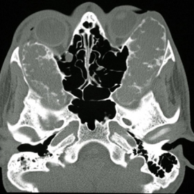
Bone window axial (a) and coronal (b) CT scan through the mid-orbits and maxilla demonstrating maxillary involvement and decreased bilateral orbital volume.


Anterior view of volume-rendering 3D CT scan showing gross bony hypertrophy of the orbital floors (L> R).

Axial CT imaging demonstrating multicystic bone lesions with expansion and erosion in the mandible and the maxillae.

Medical Analysis Report
I. Imaging Findings
The patient is a 12-year-old boy with a previous history of hereditary Cherubism (“cherubic face”). Based on the provided CT images and 3D reconstruction, the following features are observed:
- Bilateral jaw lesions: The mandible and maxilla exhibit multiple, septated (“multi-cystic”) changes with cystic bone destruction, thinning of the cortical bone in some areas, and local expansion.
- Maxillary involvement: The maxillary lesion is significant, involving both maxillary sinuses and leading to reduction or obliteration of the sinus cavities. There is partial destruction or involvement of the orbital floor, resulting in thinning or compression of both orbital floors.
- Facial asymmetry or morphological changes: On 3D reconstruction, there is fullness of the cheeks, particularly noticeable in the lower and mid-face. Axial and coronal images show upward displacement of the orbital contents, consistent with proptosis (exophthalmos).
- Periorbital changes: Involvement of the orbital floor and lateral wall may cause upward or forward displacement of the eyeball, potentially related to bone overgrowth and expansion of the lesion.
II. Differential Diagnosis
Based on the above imaging findings and the patient's history of “Cherubism,” Cherubism should be primarily considered. At the same time, multiple cystic lesions and bone overgrowth in the mandible and maxilla could also arise from the following conditions, though they are less likely compared to the previously confirmed diagnosis:
- Familial Giant Cell Granuloma: More common in younger individuals but usually presents unilaterally, making bilateral involvement rare.
- Fibrous Dysplasia: Can show a “ground-glass” appearance on imaging and typically appears in childhood or adolescence. When lesions are expansive, they may resemble Cherubism radiologically.
- Aneurysmal Bone Cyst (ABC): May present with multiple cystic radiolucencies, but pathologically features blood-filled cavernous spaces, rather than the typical multi-loculated bony structure characteristic of Cherubism.
- Other jaw cysts or tumors (e.g., ameloblastoma, odontogenic cysts): Generally localized or unilateral, which does not fit the bilateral symmetrical pattern.
III. Final Diagnosis
Taking into account the patient’s age (12 years), the previously confirmed diagnosis of Cherubism, clinical manifestations (progressive facial swelling and eye protrusion), as well as the typical findings on imaging of bilateral, multicystic bone destruction and expansion involving the orbital floor, these strongly suggest Cherubism. Hence, the most likely final diagnosis is:
Cherubism (“Cherubic Face”)
IV. Treatment and Rehabilitation Plan
1. Treatment Strategies
- Conservative observation: For early or relatively stable lesions, periodic follow-up and imaging are recommended. In childhood and adolescence, Cherubism often stabilizes or partially regresses after puberty.
- Surgical treatment: In cases of significant deformity or serious orbital/optic nerve compression leading to visual impairment or reduced quality of life, surgical intervention (e.g., curettage of the lesion, bone contouring, orbital floor reconstruction) may be considered. The timing should be individualized based on skeletal maturity to avoid adverse effects on normal growth.
- Pharmacotherapy: Currently, there is no specific medication for Cherubism. A few cases have explored the use of bisphosphonates to control bone resorption, but further evaluation of efficacy is needed.
2. Rehabilitation / Exercise Prescription
Because lesions in the jaw and orbital floor can change facial appearance and eye function, rehabilitation should be comprehensive, covering both skeletal status and general fitness principles.
- Basic phase (FITT-VP principle):
- Frequency: 3–4 times per week.
- Intensity: Low to moderate, avoiding impact or trauma to the face.
- Time: 20–30 minutes per session, gradually extending to 30–40 minutes depending on tolerance.
- Type: Activities such as walking, slow jogging, or swimming that minimize vibration of the facial region; avoid high-impact sports.
- Progression: As the child’s fitness and skeletal development progress, exercise intensity can be gradually increased. Any increase should be incremental.
- Precautions:
- Avoid direct pressure or impact to the maxillofacial area (use facial protection in ball sports, for example).
- Monitor for vision changes or worsening proptosis; seek medical attention if these occur.
- After surgery, follow specialized rehabilitation instructions, including localized exercises that facilitate the restoration of bony structures.
These rehabilitation principles are for general reference and should be tailored to the individual’s condition (bone stability, severity of orbital involvement, need for surgery, etc.). Before trying new forms of exercise or increasing intensity, consult a professional physician or rehabilitation therapist.
Disclaimer: The above report is for reference only and cannot replace an in-person consultation or professional medical advice. For a more definitive diagnosis or treatment, please consult a specialist or undergo further imaging, laboratory, and pathological examinations.
Human Doctor Final Diagnosis
Orbital involvement in cherubism