


We present the case of a 74-year-old male patient with central paresis of the right peroneal nerve.
The patient had a history of left hemicolectomy 4 months previously due to intestinal ischemia from inferior mesenteric artery thrombosis. The severity of his post-surgical course had forced him into prolonged immobilization during which he had suffered from multiple decubitus ulcers, mostly in the sacral region, and atrophy of the gastrocnemius and the quadriceps muscles. An EMG was performed and revealed central paresis of the right peroneal nerve. CT examination of the abdomen performed at that time demonstrated bilateral, linear calcifications of the gluteal muscles, more prominent on the right, adjacent to the anatomic site of the sacral nerve.
The course of the sacral nerve is as folows: it exits from the pelvis through the subpiriform fissure and passes from the posterior surface of the internal thyroid muscle. It consists of two major trunks, the tibial nerve and the common peroneal nerve, which are surrounded by a common sheath, and they divide into two separate parts at a different anatomic height in each individual. In case of a division at the level of the pelvis, the peroneal nerve exits through the piriform muscle. Myositis ossificans (MO) is a condition characterized by focal, benign and self-limiting heterotopic bone formation typically occuring within the skeletal muscle or soft tissue with unknown pathogenesis. It is most often caused by trauma (MO traumatica or circumscripta), or other froms of tissue injury (surgical procedures or prolonged immobility). In these cases, hematoma formation, tissue injury and anoxia are possible predisposing factors. Sometimes MO may develop without known trauma. In addition, there is another form, Fibrodysplasia ossificans progressiva , which is a very rare autosomal dominant genetic disorder where the ectopic ossification begins in early childhood. It is characterized by episodes of permanent heterotopic ossification of soft tissues, occurs worldwide without racial, ethnic, or geographic predilection. There is no effective treatment, and soft-tissue trauma (eg, biopsies, surgical procedures, intramuscular injections, or mandibular blocks for dental procedures) and viral illnesses are likely to induce episodes of rapidly progressive heterotopic ossification, with resultant permanent loss of motion in the affected area. Favourite locations of MO development are the extremities, particularly the thigh, hip, elbow, shoulder and knee, although unusual locations such as the paravertebral muscles have been reported. The most frequently occurring symptoms are pain, tenderness and the presence of a soft tissue mass; however the lesion may be an incidental finding. Local excision is generally curative. On CT scans, MO usually appears as a ring-like structure with a variable degree of calcification or ossification. Its center is isodense or hypodense as compared to the surrounding muscle. The degree of peripheral calcification continuously increases. The calcified structures may appear as being punctiform or cloudy, but are subtly radial. They may partially or completely fill the mass. There are no specific imaging findings for MO and by using imaging alone it is usually difficult to differentiate between the different types of MO and other pathologies, such as inflammatory lesions and neoplasms. However, the imaging history, for example a previously demonstrated hematoma on US, may indicate the traumatic form, and the presence of lesions in a child which progressively deteriorate and expand, the heritable form. The differential diagnosis of MO mostly includes the possibilities of extraskeletal or juxtacortical osteosarcoma but also extraskeletal osteochondroma, chondrosarcoma, reactive periostitis (when the MO is located directly in the periosteum), tumoral calcinosis etc. Despite advances in medical imaging, it remains difficult to distinguish MO from other disorders. As a consequence, a biopsy is frequently performed and is sometimes essential for the final diagnosis.
Myositis ossificans of the gluteal muscles.


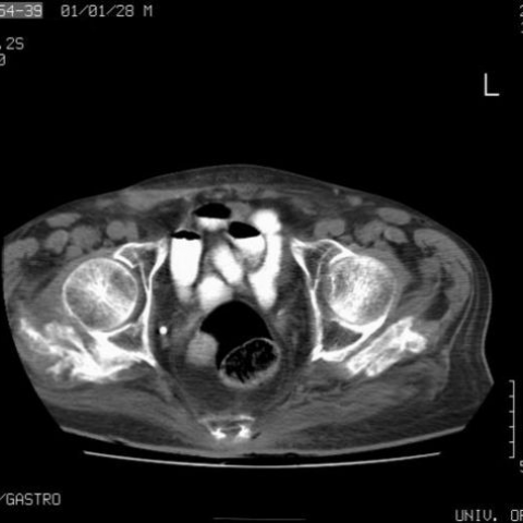

Based on the provided X-ray and CT images, a focal soft tissue mass can be observed in the anterior pelvis and pelvic cavity. Within the lesion, ring-like or irregular calcification/ossification can be seen, appearing as relatively higher density at the periphery, while the center shows iso- or low-density areas. Compared with surrounding skeletal structures, the lesion margins display new bone or calcification features. Although the lesion exerts some pressure on surrounding soft tissues, there are no obvious signs of bone destruction. Considering the patient's lower limb neurological symptoms (mild palsy of the right common peroneal nerve), it is inferred that adjacent tissues, muscles, or nerve pathways may be affected.
Taking into account the patient’s advanced age, mild palsy of the right common peroneal nerve, a focal ring-like ossification in imaging, and no obvious destruction of adjacent bone, along with the lesion morphology resembling a typical intramuscular calcified focus, the most likely clinical diagnosis is Myositis Ossificans. Nonetheless, if there is concern for a tumor or significant growth in the lesion, a biopsy and pathological examination might be warranted to rule out malignancy or other less common conditions.
1. Treatment Strategy
2. Rehabilitation and Exercise Prescription (FITT-VP Principle)
In practical application, particular attention must be paid to the patient’s overall physical condition. If there are comorbidities such as poor cardiopulmonary function or low bone density, the rehabilitation and exercise plan should be adapted under the guidance of a qualified rehabilitation practitioner or physician to ensure safety.
This report is a reference analysis based on current imaging findings and patient history. It does not replace in-person diagnosis or a professional medical consultation. If the condition changes or if there are any concerns, please seek medical attention, or proceed with further examinations and treatment under specialist guidance.
Myositis ossificans of the gluteal muscles.