Rectus sheath haematoma


Clinical History
85-year-old female under anticoagulation therapy admitted with acute onset abdominal pain and underwent ultrasound and computed tomography examination.
Imaging Findings
An 85-year-old female suffered from Alzheimer disease, with a history of congestive heart failure and atrial fibrillation was under long-term anticoagulation. A history of right nephrectomy several years ago was also referred from the relatives. The patient admitted with non-specific acute onset abdominal pain. On physical examination of the abdomen there was an abdominal tenderness at the left upper quadrant. Abdominal ultrasound confirmed at longitudinal scans a biconcave mass in the abdominal wall (Figure 1) and at transverse scans an ovoid lesion. The lesion was inhomogeneous and within it a hematocrit sign visualized at three separate foci.
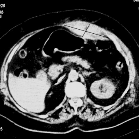
A thin line indicating the cellular component and stayed at the boundary between the plasma and cellular components in liquefied hematoma. This appearance was compatible with haematoma of rectus sheath, above the arcuate line. Due to previous history of renal cancer the patient underwent a computed tomography examination. Pre-contrast CT scans (Figure 2) revealed a biconcave hyperdense lesion within the anterior abdominal wall at the left side and a post-contrast scan (Figure 3) revealed no contrast medium enhancement of the lesion. The diagnosis of rectus sheath haematoma was confirmed. Anticoagulant therapy was stopped and she improved with conservative management.
Discussion
Rectus sheath haematoma (RSH) [1,2,3,4] is a well- described entity and an unusual cause of acute abdominal pain [2], with a reported incidence of misdiagnosis as high as 93%. In one series of ultrasound for abdominal pain, 1.8% had RSH [2]. RSH occurs 2 to 3 times more often in women [1,2,3 ] that in men. The higher incidence in women is presumably due to decreased muscle mass as compared with men. It can be traumatic or spontaneously.
Predisposing factors[1,2,3], include anticoagulant therapy, trauma, coughing, straining, pregnancy, blood dyscrasia, degenerative muscular disease, asthmatic attacks, bronchitis or influenza. High number of patients who developed RSH was under anticoagulation. The cause of RSH is the rupture of epigastric vessels [1,2,3,4].
The clinical manifestation is that of a sudden abdominal pain severe on movement. Depending on the size it may causes hemodynamic instability and symptoms and signs secondary to compression of underlying organs. Abdominal examination usually demonstrates a palpable mass, but in about a half of the cases a palpable mass is missing as it was in our case.
Plain X-ray is not specific. Ultrasound [4] is the usual first line examination with reported sensitivity 85-96% but at times can be misleading and the diagnosis established by Computed tomography examination and regarded as the examination of choice with 100% sensitivity. In subacute cases, magnetic resonance may be required for proper diagnosis.
The differential diagnoses include several entities[1,2,3,4] such as appendicitis, incarcerated inguinal hernias, urinary obstruction, acute cholecystitis, mesenteric vascular insult, dissecting aneurysms, torsion of ovarian cyst, ovarian tumor. This is because of the absence of the posterior layer of the rectus sheath below the arcuate line, allowing the haematoma to assume enormous size and be mistaken for another entity.
The patient treated conservatively with bed rest, analgesia, ice packs and haematoma compression. In cases of moderate hematocrit dropped blood or platelet transfusion needed and in severe cases surgical evacuation with ligation of bleeding vessel.
Differential Diagnosis List
Final Diagnosis
Rectus sheath hematoma due to anticoagulant therapy
Liscense
Figures
Abdominal ultrasound

Abdominal ultrasound

Addominal CT examination.
Imaging Findings
The patient is an 85-year-old female who presented with acute abdominal pain and has a long-term history of anticoagulant therapy. Ultrasound and CT examinations indicate an irregular fluid density or slightly mixed density lesion near the midline of the abdominal wall, with an abnormal appearance of the local muscle layer structure. The boundary is relatively clear or partially indistinct. On ultrasound, the lesion appears as a localized hypoechoic or mixed echo area, while on CT, it manifests as an irregular area of high, iso, or slightly low density within or anterior/posterior to the abdominal wall muscle layer, which is consistent with a hematoma. No obvious signs of organic lesions in the abdominal solid organs were observed.
Potential Diagnoses
- Rectus Sheath Hematoma (RSH)
Based on the patient’s advanced age, history of anticoagulant therapy, and imaging findings, this diagnosis is most consistent. The sudden onset of localized pain, with or without a palpable mass, and imaging showing a localized hematoma in the abdominal wall support this diagnosis. - Other Possible Acute Abdominal Conditions
These include appendicitis, torsion of an ovarian cyst pedicle, incarcerated inguinal hernia, cholecystitis, mesenteric vascular events, and others. However, these conditions typically exhibit characteristic imaging findings involving the affected organ or local structures, which were not observed in this patient’s ultrasound or CT scans; thus, they are relatively unlikely.
Final Diagnosis
Considering the patient’s age, symptoms, clinical history (especially the background of anticoagulant therapy), imaging characteristics, and clinical examination results, the most likely diagnosis is: Rectus Sheath Hematoma (RSH). If further exclusion of other rare abdominal wall or intra-abdominal pathologies is necessary, repeat ultrasound, symptomatic treatment, and close follow-up may be considered for comprehensive assessment. For cases where the diagnosis remains difficult, MRI can be employed to further clarify the presence and extent of the hematoma.
Treatment Plan and Rehabilitation
Treatment Strategies:
1. Conservative Treatment: For hemodynamically stable patients with smaller hematomas, absolute or relative bed rest, local pain management, ice packs, and compression bandaging can be used to prevent further bleeding. Adjusting or suspending anticoagulant therapy as appropriate and closely monitoring hemoglobin levels and coagulation function are recommended.
2. Symptomatic Treatment: If hemoglobin levels drop significantly, transfusion of red blood cells or platelets may be considered. Intravenous hemostatic agents can be administered if necessary.
3. Surgical Intervention: For patients with a large bleeding volume, extensive hematomas, and hemodynamic instability, urgent surgery or interventional embolization is required to remove the hematoma and ligate or embolize the bleeding vessel.
Rehabilitation/Exercise Prescription:
▪ Due to the patient’s advanced age and recent abdominal wall hematoma, it is essential to strictly limit strenuous activities in the early phase and avoid stretching or repetitive weight-bearing movements.
▪ Early Stage (e.g., within 1–2 weeks after the hematoma stabilizes): Focus on isometric abdominal muscle contraction exercises to avoid excessive stress on the abdominal muscles. Slight leg raises or abdominal contractions can be attempted in a seated or supine position 1–2 times a day for about 5 minutes each time, gradually increasing the frequency based on the patient’s tolerance.
▪ Intermediate Stage (2–6 weeks): If pain has subsided and the hematoma has significantly resolved, gradually strengthen core muscles with exercises such as supine head lifts and bridge training. Emphasize a small range of motion and slow speed, with each session lasting 5–10 minutes, 3–4 times per week.
▪ Later Stage (6 weeks and beyond): Gradually resume daily activities and light physical exertion, including walking or gentle stretching exercises as appropriate. Progressively enhance abdominal muscles and core stability, while continuing to avoid movements that markedly increase intra-abdominal pressure.
▪ Adjust the duration and intensity of exercises according to the FIT(T)-VP principle (Frequency, Intensity, Time, Type, Progression), individualizing the plan and making timely modifications based on symptom changes. If significant discomfort or increased pain occurs, seek medical evaluation promptly.
Disclaimer
This report is based on the preliminary analysis of the available imaging and clinical information. Its content is for reference only and cannot replace in-person consultations or professional medical advice. If you have any questions or if your symptoms worsen, please consult a qualified physician as soon as possible for further evaluation.
Human Doctor Final Diagnosis
Rectus sheath hematoma due to anticoagulant therapy