Spontaneous osteonecrosis of knee


Clinical History
A 46-year old male patient with three-week history of right knee pain
Imaging Findings
A 46-year-old fit active male presented with three-week history of right knee pain. No history of direct trauma to the knee. There was no previous history of knee problems to note.
On clinical examination knee is slightly swollen with tenderness over the medial femoral region. Both flexion and extension are slightly restricted.
Image findings:
Plain radiographs were within normal limits. MRI showed joint effusion, medial femoral condyle bone marrow oedema with subchondral linear low signal area consistent with fracture and fibrillation of the articular cartilage of the medial compartment of the joint. Appearances are in keeping with spontaneous osteonecrosis of knee (SONK).
Discussion
Spontaneous osteonecrosis of the knee (SONK) usually presents with acute onset of knee pain despite the patient having no significant history of trauma. It is most commonly seen in the 5th - 7th decade with a male to female ratio of 1:3. The weight-bearing portion of the medial femoral condyle is the most frequent location. However, it has also been reported in the lateral femoral condyle and both tibial plateau. SONK is often found in association with meniscal tears, following prior arthroscopy or meniscal resection, and following a diagnosis of chondromalacia associated with osteoarthritis.
Plain radiographs in the initial stages appear normal. Flattening of the weight bearing surface, subchondral sclerosis and collapse eventually lead to secondary osteoarthritis in the later stages. Triple phase isotope bone scans show increased uptake at the site of lesion. MRI is more sensitive than bone scanning and plain radiography. It provides earlier and more extensive information on the distribution of marrow abnormalities and the presence of cartilage damage, which is relevant to prognosis. Bone marrow oedema (BMO) and subsequent necrosis change the normal fat signal of the marrow with the classical appearance of hypointense regions on T1 and hyperintense regions on T2-weighted sequences. SONK can be further associated with insufficiency fracture. An insufficiency fracture is seen on MRI as subchondral, crescentic or linear dark signal regions on both T1 and T2 weighted images.
A pattern of BMO isolated to one side of a weight bearing articulation in the lower extremity has previously been attributed to many different conditions such as, transient osteoporosis, transient bone marrow oedema syndrome, true osteonecrosis, spontaneous osteonecrosis and shifting bone marrow oedema. The differentiation of these conditions is often difficult and confusing due to overlapping radiological findings and identical clinical presentations. Currently the most accepted proposed etiology is that the weight bearing articular surface is subject to altered stresses which predisposes to the development of insufficiency fractures. Some of the conditions such as transient osteoporosis and transient bone marrow oedema syndrome are merely early manifestations of subchondral insufficiency fractures. Those cases where these fractures fail to heal in the early stages are thought to progresses further to osteonecrosis.
Treatment of SONK is mainly conservative including non-steroidal anti-inflammatory drugs, protected weight bearing with crutches and physiotherapy. Surgical intervention has been shown to have mixed results
Differential Diagnosis List
Final Diagnosis
Spontaneous osteonecrosis of knee(SONK)
Liscense
Figures
plain radiograph
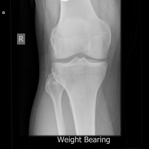
plain radiograph

Coronal PD fat saturated image

Sagittal T1w Image

Imaging Findings
1. From the right knee X-ray (including AP and lateral views), subtle variations in the bone trabeculae can be observed on the weight-bearing surface of the medial femoral condyle, showing slight sclerosis or possibly minor indentation.
2. On the MRI coronal and sagittal sequences, the medial femoral condyle reveals localized bone marrow signal alterations: hypointense on T1-weighted images and hyperintense on T2-weighted images, suggesting bone marrow edema (BMO).
3. The MRI shows a band-like or crescent-shaped hypointense area beneath the weight-bearing surface of the medial femoral condyle, indicative of a subchondral fracture line (ischemic or micro-fracture line), consistent with common features of spontaneous osteonecrosis of the knee (SONK) or a subchondral fracture.
4. There is no definite large area of cartilage collapse or severe destruction seen at this stage; however, further follow-up is recommended to monitor possible cartilage injury or secondary osteoarthritis changes.
Potential Diagnoses
Based on the patient’s age, symptoms, and imaging findings, the following diagnoses or differentials may be considered:
1. Spontaneous Osteonecrosis of the Knee (SONK): Commonly seen in middle-aged and older patients, often involving the weight-bearing surface of the medial femoral condyle. MRI typically shows localized bone marrow edema and osteonecrotic changes, sometimes with a subchondral fracture line.
2. Subchondral Fracture/Insufficiency Fracture: Frequently occurs in areas with weakened bone or increased local stress, presenting as a crescent-shaped hypointense line and bone marrow edema on MRI.
3. Early Osteoarthritis (OA): Common in middle-aged and older patients, often seen on X-ray as joint space narrowing and osteophyte formation. In mild or early stages, it may be indistinguishable from other conditions.
4. Transient Bone Marrow Edema Syndrome: Clinically and radiologically manifests as pronounced bone marrow edema, typically resolving over weeks to months. Although more common in the hip joint, it can occur in the knee and should be included in the differential diagnosis.
Final Diagnosis
Taking into account the patient’s age of 46, acute onset of right knee pain, the absence of severe destructive changes on X-ray, and the characteristic bone marrow edema with a crescent-shaped lesion on MRI, the most likely diagnosis is:
Spontaneous Osteonecrosis of the Knee (SONK) with evidence of a subchondral fracture (sub-fracture line).
If uncertainty remains, further follow-up or additional MRI examinations, or arthroscopic evaluation, may be considered to clarify cartilage integrity and potential meniscal lesions.
Treatment and Rehabilitation Plan
1. Conservative Treatment
(1) Nonsteroidal Anti-inflammatory Drugs (NSAIDs): To alleviate joint pain and inflammation.
(2) Weight-Bearing Restriction: Use of a cane or a walker is advised to reduce load on the joint and prevent fracture propagation or collapse.
(3) Physical Therapy: Heat therapy, ultrasound, or low-frequency electrical therapy to enhance local circulation and relieve pain and muscle tension.
2. Surgical Intervention
If conservative management fails or if there is obvious joint surface collapse, progressive worsening of pain, or concurrent joint pathology (e.g., meniscal tear), options such as arthroscopic surgery or orthopedic procedures (e.g., osteotomy, joint replacement) may be considered. The choice depends on the extent of the lesion and the patient’s functional requirements.
3. Rehabilitation/Exercise Prescription
Follow the FITT-VP principle (Frequency, Intensity, Time, Type, Progression, and Individualization):
(1) Early Stage (Acute Pain Phase):
• Frequency: 2–3 times a day of light exercise (e.g., straight leg raises, ankle pumps), around 10 minutes each session.
• Intensity: Avoid or minimize weight-bearing activities, focusing on non-weight-bearing or minimal weight-bearing exercises.
• Time: Adjust according to tolerance, gradually increasing exercise duration.
• Type: Engage in lower-limb active movements while lying in bed or sitting, avoiding forceful knee flexion and extension.
(2) Middle Stage (Stable Symptoms Phase):
• Frequency: 3–4 times per week, gradually increasing exercise variety and sets.
• Intensity: Introduce mild weight-bearing training (e.g., short-distance walking on flat ground) and strengthen periarticular muscles (quadriceps, hamstrings).
• Time: 15–20 minutes per session, adjusted based on pain and fatigue level.
• Type: Carefully perform closed-chain exercises (e.g., seated leg presses) or low-resistance cycling.
(3) Late Stage (Functional Recovery Phase):
• Frequency: 3–5 times per week, progressively returning to daily weight-bearing activities.
• Intensity: Increase weight-bearing tasks as tolerated without significant pain, such as slow uphill walking, stair training, and continued strengthening of knee-supporting muscles for stability.
• Time: 20–30 minutes each session or longer, depending on the individual’s condition.
• Type: Incorporate flexibility training, balance exercises, and light aerobic activities (e.g., swimming, elliptical) under safe conditions.
4. Special Considerations
(1) Throughout the rehabilitation, closely monitor knee pain, swelling, and functional status. If symptoms worsen, seek medical evaluation promptly.
(2) If the patient is at risk of osteoporosis or has insufficient cardiopulmonary function, coordinate with specialists to tailor training intensity and methods accordingly.
Disclaimer
This report is solely based on the provided imaging and clinical information for reference and does not substitute for an in-person consultation or professional physician’s diagnosis and treatment. If you have further questions or changes in your condition, please consult an orthopedic or radiology specialist promptly.
Human Doctor Final Diagnosis
Spontaneous osteonecrosis of knee(SONK)