Unstable osteochondritis dissecans


Clinical History
Three weeks of pain in the knee on medial femoral condyle. It was associated with a traumatic event.
Imaging Findings
Three weeks of pain in the knee on medial femoral condyle. It was associated with a traumatic event. His general practitioner suggested non-steroidal anti-inflammatory medication along with physiotherapy, but there was no improvement.
The AP radiograph was negative.
The MR imaging protocol included gradient T2 weighted in coronal sagittal and axial plain. The images showed small displaced bone fragment of the medial femoral condyle, like unstable lesion.
There was presence and size of a high-signal-intensity line between the osteochondritis dissecans fragment and the underlying bone. It was osteochondritis dissecans.
Discussion
Osteochondritis dissecans is a form of osteochondrosis limited to the articular epiphysis. Articular epiphyses fail as a result of compression. Both trauma and ischemia probably are involved in the pathology. Trauma is most likely the primary insult, with ischemia as secondary injury. Trauma may be caused by direct trauma, such as impaction fracture, or repetitive microtrauma, such as excessive normal compressive stress.
The pathology of osteochondritis dissecans may be described in 3 stages.
In the first stage (acute injury), edematous intra-articular and periarticular soft tissues are observed. In the second stage, the epiphysis reveals a thinning of the subcortical zone of rarefaction. On radiography, the epiphysis may demonstrate fragmentation.
The third stage is the period of repair in which granulation tissue gradually replaces the necrotic tissue. Necrotic bone may lose its structural support, which results in compressing and flattening of the articular surface.
In the knee joint, the medial femoral condyle is the most commonly involved site. Potential locations are the lateral aspect of the medial femoral condyle (75%), the weight-bearing surface of the medial (10%) and lateral femoral condyles (10%), and the anterior intercondylar groove or patella (5%).
In femoral condyles, it has been estimated to occur in 6 per 10,000 men and in 3 per 10,000 women younger than 50 years. In general, osteochondritis dissecans occurs more commonly in the convex surface than in the concave surface of a joint. Since the advent of cross-sectional imaging (CT and MRI), OCD of the talus has been diagnosed more frequently and, in future series, may represent the most frequent site of OCD.
OCD tends to affect young patients, pain is the primary symptom. No racial predilection is recognized and osteoarthritis is a common long-term complication.
Patients usually report pain at the extremes of motion range. Periarticular edema is often present with slight warmth to the touch. When a lower extremity is involved, patients often limp. Symptoms usually improve with protected immobilization of the joint. On conventional radiographs osteochondral lesion may appear normal. When detectable osteochondral lesions appear as lucencies in the articular epiphysis.
MRI correlates best with surgical staging and detects occult lesions that also may not be evident on CT. A STIR sequence is the most sensitive. On T2-weighted images, a high signal intensity line in the zone, indicative of fluid or granulation tissue, has proved to be a frequent and important sign.The presence of fluid encircling the fragment or focal cystic areas beneath the fragment are the best indicators of such instability.
Now MRI is used for long-term follow-up studies to assess the condition of the bony fragment, parent bone and interface, so as to determine no change, partial or complete remission, or progression of osteochondritis dissecans.
Osteochondritis dissecans, as described in the earlier literature, usually was treated either nonoperatively or with an arthrotomy for removal of the loose fragment
Closed treatment is still preferred for patients who are first seen early in the disease process and for those who have open physes.
Differential Diagnosis List
Final Diagnosis
Instabil osteochondritis dissecans
Liscense
Figures
MR-coronal plain

MR-sagittal plain

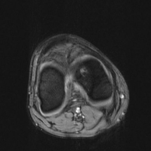
MR-axial plain
MR-coronal plain

Medical Analysis Report
I. Radiological Findings
The patient is a 14-year-old male with a chief complaint of medial knee pain for three weeks. The provided MRI images of the knee show the following main features:
- The lesion is located in the medial femoral condyle (often seen at the lateral margin of the medial femoral condyle), presenting as abnormal signals beneath the articular surface.
- Irregular subchondral bone and signal alterations are observed locally. On T2/STIR sequences, higher signal intensity may indicate bone marrow edema or fluid-like signals.
- In some slices, a low-signal band or high-signal line is visible beneath the cartilage surface, suggesting possible loosening or instability of osteochondral fragments.
- Mild surrounding soft-tissue edema signal is noted, with no obvious large effusion seen in the joint cavity.
II. Possible Diagnosis
Combining the patient's age, clinical history, and imaging findings, the following diagnoses or differential diagnoses are considered:
- Osteochondritis Dissecans (OCD): Most commonly seen in adolescents, often involving the femoral condyle. On T2-weighted images, irregular or ring-like high signals can indicate fragment mobility and instability at the bone-cartilage interface.
- Avascular Necrosis (AVN): It can present with abnormal signal changes in the distal femur, but more commonly involves collapse of the articular surface or occurs in the hip. Compared to this case, the patient’s younger age and imaging features suggest OCD is more likely.
- Other articular cartilage or subchondral bone lesions: Such as subchondral cysts or microfractures. However, based on the imaging findings and clinical history of this case, these are less likely.
III. Final Diagnosis
Considering the patient’s typical age (adolescence), clinical symptoms (knee pain), and imaging changes (osteochondral separation-like changes and a potential instability line in the medial femoral condyle), the most likely diagnosis is: Osteochondritis Dissecans (OCD).
If clinical symptoms worsen or if there is poor response to treatment, further MRI follow-up and arthroscopic evaluation may be considered. Biopsy or more definitive procedures may be needed for further confirmation.
IV. Treatment Plan and Rehabilitation
1. Conservative Treatment:
- In early stages or in adolescents with open growth plates, weight-bearing restriction, bracing, or using crutches to reduce stress may be employed.
- Anti-inflammatory and analgesic medications (e.g., NSAIDs) can be used to relieve pain and inflammation.
- Regular outpatient follow-ups and repeat imaging are recommended to assess disease progression.
2. Surgical Treatment:
- If there are clearly loose or detached osteochondral fragments, unstable articular surfaces, ineffective conservative treatment, or severe lesions, surgical intervention such as arthroscopic fixation, drilling decompression, or grafting may be recommended.
- Postoperative rehabilitation exercises are necessary to restore joint function and prevent re-injury.
3. Rehabilitation/Exercise Prescription Recommendations (FITT-VP Principle):
- Frequency: Initially, exercises targeting the lower limbs (e.g., isometric and isotonic training for the quadriceps) can be performed 3–5 times per week, adjusted according to pain levels.
- Intensity: Begin with low-intensity, closed-chain exercises such as seated knee extensions and straight-leg raises. Gradually progress; avoid significant pain during each workout.
- Time: Each session can last 20–30 minutes, and can be lengthened as tolerated. Multiple short breaks are possible as needed.
- Type: Progress from non-weight-bearing or partial-weight-bearing exercises to weight-bearing exercises, then gradually incorporate proprioception training and low-impact aerobic activities (e.g., swimming, stationary cycling).
- Progression: Increase difficulty and loading gradually according to pain relief and muscle strength recovery. As follow-up imaging shows improvement, running and jumping activities can be resumed stepwise.
Disclaimer:
This report is based solely on the provided medical history and imaging information for reference and cannot replace an in-person consultation or professional medical diagnosis and treatment. Specific treatment plans must be determined comprehensively according to the patient's actual condition, further examination results, and specialist opinions.
Human Doctor Final Diagnosis
Instabil osteochondritis dissecans