Segond fracture and tibial spine avulsion fracture secondary to sporting injury


Clinical History
Segond fracture and tibial spine avulsion fracture secondary to twisting injury of knee.
Imaging Findings
A 13 year old male patient sustained the above injury when his leg twisted awkwardly under his motorbike after having fallen off during a motor-cross race.
His knee was immediately painful, swollen and he was not able to weight bear. Clinical examination revealed a large effusion of the knee, tenderness laterally and no distal neurovascular deficit. Plain radiographic examination demonstrated a lateral avulsion fracture just distal to the tibial plateau, consistent with a Segond fracture. Interestingly, the radiograph also demonstrated an avulsion of the tibial spine, consistent with an anterior cruciate ligament injury, of which a Segond fracture is virtually pathognomic.
Open reduction and internal fixation of the tibial spine avulsion fracture was performed via a midline approach. The tibial spine was relocated into position and secured with a 30x4.5mm, partially threaded cannulated screw. Post operatively the patient was mobilised non weight-bearing in a hinged knee brace.
Discussion
A Segond fracture is a vertical avulsion fracture of the proximal tibia, just distal to the tibial plateau. It was first described by Dr Paul Segond in 1879 as a small avulsion fracture off the lateral tibial plateau at the insertion of the mid-portion of the lateral collateral ligament, posterior to Gerdy’s tubercle. It is sustained by forced internal rotation and a varus stress which places abnormal stress on the lateral collateral ligament. The pathogenesis is related to the attachment of the anterior oblique band of the lateral collateral ligament and ilio-tibial band fibres to the avulsed fragment. The importance of recognition of a Segond fracture is that the severity of the mechanism of the injury causes injury to the anterior cruciate ligament and in most cases also a meniscal injury or damage to the posterolateral corner of the knee.
The lateral avulsion fracture can be seen clearly on the anteroposterior plain radiograph whilst the avulsed tibial spine is clearly seen on both lateral and anteroposterior views.
In children it is common for an ACL injury to present as an tibial spine avulstion fracture. In contrast, often in adults a Segond fracture will be observed with an absence of a tibial spine fracture due to the relative reduced strength in the anterior cruciate ligament.
The current case clearly demonstrates the relation between lateral collateral and anterior cruciate ligament injuries as seen on plain radiograph (see figure 1).
Imaging procedures together with clinical history and examination confirmed the diagnosis of a Segond fracture and a comminuted fracture of the tibial spine (see figure 2).
MR scan can be useful preoperatively to further assess the extent of injury. In the current case this was not performed as open surgery was planned for fixation of the tibial spine avulsion fracture. Examination under anaesthetic and direct visualisation of the menisci at that stage confirmed the abscence of further soft tissue injury.
Differential Diagnosis List
Final Diagnosis
Segond fracture and tibial spine avulsion fracture
Liscense
Figures
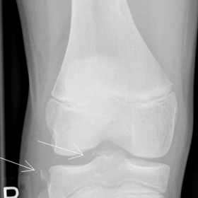
Plain radiograph


CT scan


Imaging Findings
Based on the provided X-ray and CT images, the following findings are observed:
1. A small avulsed bone fragment on the lateral tibial plateau (near the attachment site of the lateral collateral ligament), consistent with the typical characteristics of a Segond fracture. This fracture usually occurs at the lateral margin of the proximal tibia, just posterior to Gerdy’s tubercle, and may present as a vertically oriented small avulsion fragment.
2. Multiple fragments at the intercondylar eminence (tibial spine), showing a comminuted pattern, consistent with a tibial spine avulsion fracture. Both sagittal and frontal views reveal bony disruption and displacement in this area.
3. No obvious narrowing of the joint space is noted; knee joint alignment is generally acceptable, and the cortical bone of the femur and other parts of the tibia appears largely intact.
4. No evident soft tissue swelling or abnormal fluid collection is seen on imaging (bearing in mind that plain radiographs and CT have limited soft tissue resolution, so ligament or meniscal injuries cannot be completely ruled out).
Combined with clinical data, these findings suggest an avulsion fracture at the attachment site of the lateral collateral ligament of the knee, accompanied by a tibial spine avulsion fracture. The imaging indicates a relatively severe twisting injury with valgus and internal rotation stress, calling for special attention and evaluation of the anterior cruciate ligament (ACL), the lateral collateral ligament, and other important soft tissue structures.
Potential Diagnoses
1. Segond Fracture (Lateral Avulsion Fracture):
· Typically an avulsion fracture on the lateral proximal tibia near the knee, often associated with ACL injuries.
· In this case, the lateral avulsed fragment is clearly visible on the X-ray, consistent with a Segond fracture.
2. Tibial Spine Avulsion Fracture:
· In children and adolescents, the growth plate at the ACL attachment is relatively weak, often resulting in a tibial spine avulsion fracture rather than a ligament tear.
· In this case, a distinct avulsion fragment in the tibial spine region is present, consistent with an avulsion fracture.
3. Associated Soft Tissue/Ligament Injuries (to rule out meniscal or other major ligament tears):
· A Segond fracture often indicates stress-related injuries to the ACL and lateral collateral ligament.
· Clinically, MRI or surgical exploration may be needed to confirm the status of the meniscus and ligaments.
Final Diagnosis
Considering the patient’s age of 13, the twisting injury, and the findings on X-ray and CT—along with known associations between Segond fractures, ACL injuries, and tibial spine avulsion fractures—the most likely diagnosis is:
Segond fracture combined with a comminuted tibial spine avulsion fracture.
Surgical exploration confirmed no significant injury to the meniscus or other soft tissue, consistent with the fact that in pediatric populations, the injury commonly manifests as bone fractures rather than complete ligament tears.
Treatment Plan and Rehabilitation Protocol
1. Treatment Plan:
· Surgical Intervention: Given the comminuted tibial spine avulsion affecting the stability of the ACL attachment, internal fixation or repair is typically required. Because the growth plates are still open in children, surgical methods must be carefully selected after thorough evaluation.
· Ligament and Lateral Collateral Ligament Assessment: If significant damage to the lateral collateral ligament or soft tissues is identified intraoperatively, concurrent repair or reconstruction is considered. If the ligament remains attached to the avulsed bone fragment, fixation and reduction of the fracture may suffice for ligament healing.
· Perioperative Management: Control pain and actively prevent deep vein thrombosis. A brace may be used to maintain knee stability as needed.
2. Rehabilitation and Exercise Prescription:
(1) Early Stage (0–2 weeks post-op):
· Use of a knee brace or cast to minimize weight-bearing.
· Under the guidance of a physician or physical therapist, perform isometric quadriceps exercises and gentle ankle movements to promote circulation.
· Elevate the injured limb and apply ice as needed to alleviate pain and swelling.
(2) Middle Stage (2–6 weeks post-op):
· As permitted by the physician, gradually increase knee range of motion with passive and active flexion-extension exercises, avoiding excessive force.
· Initiate partial weight-bearing, for example, walking with crutches while tolerating pain and ensuring satisfactory fracture healing progress.
· Perform light resistance exercises for the quadriceps and hamstrings to maintain muscle strength and joint stability.
(3) Late Stage (6 weeks post-op and beyond):
· Transition to full weight-bearing as tolerated, with a progressive strengthening regimen under professional guidance. This includes seated and standing resistance exercises and core muscle training.
· Gradually introduce proprioception and stability training (e.g., balance board exercises, mild hopping drills), following a step-by-step progression.
· Depending on the recovery of fracture healing and soft tissue, progressively return to running, mild directional changes, and relevant sports activities.
(4) FITT-VP Principle:
· Frequency: Rehabilitation exercises 3–4 times per week. In the early phase, sessions can be more frequent but shorter in duration.
· Intensity: Within a pain-free or mildly uncomfortable range; increase resistance and range of motion gradually according to post-operative healing.
· Time: Begin with 20–30 minutes per session, then increase to 40–60 minutes in later phases.
· Type: Emphasize quadriceps strengthening, core stabilization, and balance/coordination training, supplemented with low-impact aerobic exercises (e.g., cycling, elliptical).
· Progression: Gradually increase weight-bearing, resistance, range of motion, and complexity of training exercises based on clinical recovery.
Disclaimer
This report provides a reference analysis based on current clinical and imaging data. Specific treatment plans should be derived from the actual clinical situation by the attending physician and the professional medical team. If you experience any discomfort or have questions, please seek immediate medical attention and consult a healthcare professional.
Human Doctor Final Diagnosis
Segond fracture and tibial spine avulsion fracture