Sequential stress fractures of the metatarsal bones: X-ray, CT and MR findings.


Clinical History
The patient presented with metatarsalgia, especially at weight bearing. There was no history of trauma.
Imaging Findings
The patient presented with metatarsalgia lasting for one month. The pain was more relevant at weight bearing. There was no history of direct trauma. The patient's investigation was negative for any bone metabolic disease.
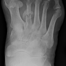
The patient underwent X-ray that showed a fracture of the second metatarsal bone; she was treated with partial weight bearing.
After 2 months, due to the persistence of pain, the patient underwent a CT examination which confirmed the presence of the second metatarsal bone fracture and it also showed the presence of a fracture within the third metatarsal bone. A bone metabolism investigation was performed (negative).
A MRI examination was performed (about one month later) in order to study soft tissues of the foot and bone marrow of the metatarsal bones. SE T1 confirmed the presence of the two fracture lines and STIR showed areas of bone marrow oedema within the proximal portion of the shaft of the first and second metatarsals, within the distal portion of the shaft of the third metatarsal bone and within the head of the fourth metatarsal bone. Moreover there was diffuse oedema of soft tissues.
These clinical, X-Ray, CT and MR features were compatible with SEQUENTIAL stress fractures (unusual presentation for multiple stress fractures, which normally occur together).
No therapy was recommended (only oral FANS to relieve the pain).
Discussion
Stress fractures occur in normal or metabolically weakened bones. They are classified into two groups: those that result from prolonged cyclical mechanical stress on normal bone are called "fatigue fractures", while those that occur with physiologic stress on bones weakened by metabolic disease or radiation treatment are called "insufficiency fractures". Incidence of fatigue fractures is increasing in the population, with runners now the most commonly affected group, accounting for 72% of stress fractures. Insufficiency fractures occur more commonly in the elderly and, in particular, in oncology patients.
Among the metatarsal bones, stress fractures involving the middle and distal portions of the shaft of the second and third metatarsals are most common. Stress fractures at the base of the first or second metatarsals or affecting other metatarsal bones are less common.
The pathogenesis of stress fractures is due to a cyclic, repetitive and submaximal loading of the bone. This creates numerous microfractures especially when the duration, intensity or frequency of physical activity is rapidly increased. If this damage is not abated through adequate rest, it ultimately exceeds the reparative ability of the skeletal system and the microfractures coalesce into a stress fracture.
Running is reported to be the predominant cause of stress fractures but other factors contribute to their development such as bone composition, vascular supply, hormonal imbalances, nutritional status and inappropriate footwear.
Most stress fractures are classified as low risk. Typically, the patient experiences an insidious onset of localised pain. Some stress fractures are classified as high risk. They manifest with vague pain and can lead to a prolonged recovery because of complications with bone union.
Radiography is the first line of diagnosis. The initial appearance may be normal but in other cases the classic feature is of a lucent fracture line associated with sclerosis, benign periosteal reaction and endosteal cortical thickening.
In CT the typical appearance of a stress fracture is that of focal callus formation and endosteal thickening around a fracture site. A helpful sign for distinguishing stress fractures from pathologic fractures is the presence of an aggressive periosteal reaction.
MRI findings include decreased marrow signal on T1 sequences and increased marrow signal on T2 sequences around a fracture line. The most sensitive discriminating feature between stress and pathologic fractures is a well-defined low signal T1 weighted abnormality around a fracture indicating an underlying tumour. For a whole-body approach, bone scans and PET scans carry an advantage over CT and MRI but they are nonspecific. In the evaluation of stress injuries, a bone scan can reveal changes before radiography but offers lower resolution and specificity to differentiate stress and pathologic fractures.
Similarly, PET scans may show FDG uptake at the site of a stress fracture, potentially mistaking the presence of a metastatic focus.
Low-risk stress fractures generally are easy to recognise and are treated with 6–8 weeks of relative rest. For high-risk stress fractures an aggressive treatment is warranted and it should be the surgical reduction of the fracture followed by 6–8 weeks of postoperative immobilisation.
Differential Diagnosis List
Final Diagnosis
Sequential stress fractures of the metatarsal bones
Liscense
Figures
X-Ray

CT


MRI


Imaging Findings
Based on the provided foot X-ray, CT, and MRI images, the following characteristics can be observed:
1. X-ray plain films: A subtle radiolucent line (possibly not very obvious) is visible at the proximal or metaphyseal region of the second metatarsal, with local cortical thickening and mild sclerosis.
2. CT scans: Show evident trabecular and cortical thickening in the suspicious area, with mild local sclerosis; in some areas, a small amount of bony healing (initial callus formation) may be present.
3. MRI: On T1-weighted images, a low-signal area is observed, while corresponding T2 or STIR (water-sensitive) sequences display a high-signal edema-like shadow in the same location (indicative of bone marrow edema). Around the suspected fracture line, there is a noticeable signal change characteristic of stress fractures (commonly seen in the tibia or metatarsals). There is no significant soft tissue avulsion or obvious mass. A small amount of edema is present in the surrounding soft tissue.
Potential Diagnoses
- Stress Fracture (Fatigue Fracture or Insufficiency Fracture): Considering the patient’s advanced age and the location of symptoms in the metatarsal area, along with the absence of a clear history of major trauma, and imaging findings of cortical thickening and local sclerosis, a stress fracture is most consistent. In older patients, these fractures are often insufficiency fractures attributable to “bone fragility” or metabolic factors.
- Pathological Fracture: If there is a history of underlying tumor or a bone-destructive lesion (e.g., metastasis), the findings may present as a fracture line or abnormal local signal. However, in general, more invasive lesions or irregular bony destruction would be seen on MRI or CT.
- Osteoporotic Fracture with Minor Trauma: In older individuals, especially females with osteoporosis, low-energy fractures can occur. Imaging findings may overlap with insufficiency fractures to some extent, and the two conditions can be interrelated.
Final Diagnosis
Taking into account the patient’s age (77 years), clinical presentation (plantar weight-bearing pain), lack of a clear traumatic event, and imaging evidence of stress-related changes and sclerosis in the second metatarsal, the most likely diagnosis is:
Stress Fracture (“Insufficiency Fracture”) of the Second Metatarsal.
If there is a need to rule out potential malignant or invasive lesions, further laboratory tests (e.g., markers of bone metabolism, tumor markers) or in certain cases a bone biopsy can be considered. With clinical correlation and additional assessments, the above diagnosis can be confirmed.
Treatment and Rehabilitation Plan
Treatment and rehabilitation for a stress fracture can be divided into several components:
- Conservative Treatment: For low-risk or mild stress fractures, partial or protected weight-bearing is recommended, using braces or protective footwear. During a 6–8 week period, reduce or avoid prolonged standing, walking, as well as high-impact activities. Use of crutches or a walker may be warranted if necessary.
- Medications and Support: Pain management can include nonsteroidal anti-inflammatory drugs (NSAIDs) or other analgesics, bearing in mind potential gastrointestinal or kidney issues in the elderly. Appropriate supplementation with calcium, vitamin D, or bone-strengthening medications (e.g., bisphosphonates) may help improve bone integrity.
- Surgical Intervention: If the fracture is considered high-risk or exhibits significant instability (for example, a fracture at the base with marked displacement), internal fixation surgery may be considered. This reduces the risk of later malalignment and promotes healing. Postoperative management typically involves 6–8 weeks of partial weight-bearing or immobilization.
-
Rehabilitation and Exercise Prescription: Rehabilitation exercises should progress gradually, following the FITT-VP principles (Frequency, Intensity, Time, Type, Volume, Progression, and Principles of individualization):
• Initial Phase (0–2 weeks): Focus on static lower limb muscle strength exercises. Perform ankle joint movements and muscle contractions in a non-weight-bearing position (e.g., in bed or seated). Frequency is 1–2 sessions per day, each for 10–15 minutes.
• Mid Phase (2–6 weeks): Gradually increase short-distance walking on flat ground under protected weight-bearing conditions, starting with 5–10 minutes each time, 2–3 times a day. Adjust intensity based on pain and fracture healing. Low-impact exercises such as light cycling or underwater walking may also be introduced.
• Late Phase (after 6 weeks): If imaging and clinical evaluations suggest good fracture healing, progressively introduce weight-bearing drills (e.g., mild heel raises, ankle stability exercises with resistance bands) and gradually increase the duration and intensity of aerobic activities. Monitor foot pain and fatigue on a weekly basis, and reduce intensity or incorporate more rest days if necessary.
Disclaimer
This report is a reference medical analysis based on the provided materials and images. It cannot replace an in-person diagnosis or guidance from a professional physician. If you have any concerns or if your condition changes, please consult an orthopedics, foot and ankle surgery, or rehabilitation specialist without delay for more accurate diagnostic and treatment advice.
Human Doctor Final Diagnosis
Sequential stress fractures of the metatarsal bones