

A 41-year-old man was admitted to the emergency department after suffering a severe torsion trauma of the right knee at work. Clinical examination revealed a severely diminished range of motion, painful mobilisation of the knee and tenderness over the lateral joint space.
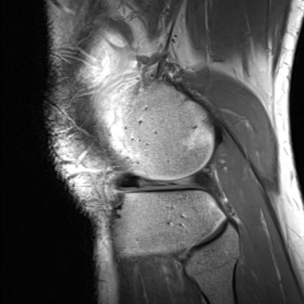
Plain radiography at the emergency department showed no abnormalities. MR imaging was performed on a 3T MRI (TrioTim Siemens, Erlangen, Germany). Sagittal proton-density and T2-weighted images demonstrated moderate hydrops. A small radial tear of the anterior horn and body of the medial meniscus was visible on the coronal and sagittal T2 images. A bucket-handle tear with complete anterior luxation of the posterior horn of the lateral meniscus was demonstrated. The absent bow tie sign and empty meniscus sign were visible on both sagittal sequences. Additional sagittal 3D proton-density sequence with fat saturation clearly demonstrated the anteriorly displaced posterior horn, located adjacent to the normal anterior horn, thus forming the double anterior horn sign. The classic double PCL sign, often visualised in medial meniscus bucket-handle tears, was not present.
The meniscus plays an important role in the biomechanics and physiology of the knee, and the absence of a normal meniscus can lead to accelerated and irreversible degenerative changes. Bucket-handle tears of the meniscus are an important and not infrequent type of meniscal injury, occurring in about 10% to 26% of meniscal tears, and are more common medially. It often affects younger individuals as a consequence of sport related trauma. Compared to other types of meniscal injury, bucket-handle tears are more common in previously normal menisci. Bucket-handle tears are longitudinal or oblique tears with an attached fragment displaced away from the meniscus. Displaced fragment are the main cause of symptoms in meniscal bucket-handle tears. It is important to diagnose meniscal tears with displaced meniscal material because they require surgery for reattachment or removal. A group of MRI signs in the diagnosis of such tears have been reported. Of these signs, double posterior cruciate ligament (PCL), fragment in the intercondylar notch, flipped meniscus, and absent tie bow sign are well known. Other signs are the double anterior horn sign (or double delta sign), the disproportional posterior horn sign, the absent meniscus sign and the coronal truncation sign. The presence of three or more of these MR imaging signs should be regarded as highly suggestive for a bucket-handle tear.
The double-PCL sign denotes a displaced meniscal fragment antero-inferior and parallel to the PCL, giving it an appearance of a double PCL. Fragment in the intercondylar notch sign defines a fragment in the intercondylar notch, like in the double-PCL sign, but not parallel to the PCL, and not in the same sagittal plane. Absent tie bow sign implies the occurrence of only one or no meniscal body segment in consecutive peripheral sagittal MR images. Flipped meniscus sign is defined as an anteriorly displaced triangular-shaped meniscal fragment located on top of the native anterior horn. This results in a seemingly enlarged anterior horn, more than 6 mm in height. If the displaced meniscal fragment is juxtaposed posterior to the native anterior horn, it is referred to as the double anterior horn sign. The disproportional posterior horn sign defines the presence of a larger posterior meniscal horn in the central sections than in the peripheral sections on sagittal MR images, and is considered to indicate a postero-centrally displaced fragment of the anterior horn. The absent meniscus sign is seen when the entire meniscus is displaced. A vertical amputation of the meniscus in the coronal plane defines the coronal truncation sign.
Anteriorly displaced bucket-handle tear of the lateral meniscus.


Based on the patient’s right knee MRI images, morphological abnormalities can be observed in the lateral meniscal area, manifested as:
Apart from this, there are no obvious signs of a complete rupture of other major ligaments (such as the anterior and posterior cruciate ligaments or collateral ligaments) visible on the current images, but partial ligament injuries should be ruled out through additional imaging sequences and physical examination.
Taking into account the patient’s mechanism of injury (twisting of the right knee), related physical findings (significant limitation in activity, tenderness in the lateral joint space), and imaging findings (abnormal meniscal morphology with visible displaced fragments), the following diagnoses or differentials may be considered:
Considering the patient’s age, twisting injury mechanism during sports/work, significant lateral joint line pain, and MRI findings indicating a longitudinal tear of the lateral meniscus with a displaced fragment, the most likely diagnosis is:
Bucket-handle tear of the lateral meniscus.
If diagnostic uncertainty persists, arthroscopic examination may be performed to definitively determine the extent of the tear and any displacement.
Rehabilitation should be performed under the guidance of a physician or physical therapist, following a progressive and personalized approach. For postoperative or acute-phase patients, the following stages may be considered:
Throughout the rehabilitation process, if the patient has bone fragility, excessive body weight, or reduced cardiopulmonary function, exercise intensity and type should be carefully selected. Knee pain and swelling should be continuously monitored. If any discomfort or exacerbation of symptoms occurs, prompt medical review is advised.
This report solely provides a reference analysis based on the available medical history and imaging data. It does not replace in-person consultation or professional clinical diagnosis and treatment recommendations. The specific treatment plan must be tailored to the patient’s actual condition and a comprehensive evaluation by a qualified physician.
Anteriorly displaced bucket-handle tear of the lateral meniscus.