Aggressive reactive periostitis (ECR 2010 Case of the Day)


Clinical History
41-year-old female violin player came with swelling and pain of her 4th finger.
Imaging Findings
Plain film manifested soft tissue swelling in the 4th finger; as well as increased density, calcifications and paraosteal-periosteal reaction of the lateral proximal phalanx (Fig. 1).
US showed calcified soft tissue mass on the transverse and longitudinal plane (Fig. 2).
On MRI an ill defined soft tissue mass was demonstrated on the volar and ulnar side of the proximal phalanx, paraosteal-periosteal irregular heterotopic calcifications were shown and intense soft tissue oedema surrounded the lesion. The cortex was intact and there was no continuity with bone marrow. On FSE T1wi lesion was iso or slightly hyperintense to muscle (Fig. 3), on fat-suppressed FSE the lesion was hyperintense (Fig. 4) and gradient echo nicely demonstrated lamellated irregular periosteal calcify mass (Fig. 5). Strong enhancement was seen after gadolinium injection (Fig. 6).
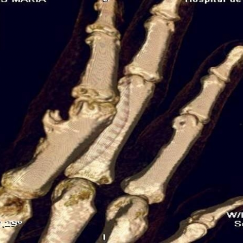
MDCT confirmed the calcifying periosteal mass proliferation without cortical involvement (Fig. 7).
Discussion
Florid reactive periostitis is an uncommon bone lesion that most often occurs in the long tubular bones of the hands and feet. It is also known as Nora’s lesion, aggressive florid periostitis or bizarre osteochondromatous proliferation (BPOP). Is part of the spectrum of reactive lesions including florid reactive periostitis and turret exostosis.
Clinically patients present with finger swelling, moderate pain and erythema associated with minimal trauma.
Its high incidence in the right hand and the predominant location on the proximal phalanx suggest traumatic or micro-trauma origin.
Radiological appearance shows bone proliferation hetero-topic ossification adjacent to the cortical bone lacking continuity with the underlying bone marrow and with intact cortex and soft tissue changes.
Pathological studies have demonstrated three different stages starting with reactive periostitis with minimal osteocartilaginous proliferation. It is followed by bone and metaplastic cartilage formation, and ends with mature ossification and cartilage cap. Radiologically these different stages can be also followed from a subtle soft tissue calcify periosteal mass to a sessile bone formation.
Its distinct radiological features allow differential diagnosis with bone tumours such as paraosteal osteosarcoma or chondrosarcoma and biopsy is usually not needed.
Differential Diagnosis List
Final Diagnosis
Aggressive reactive periostitis
Liscense
Figures
Plain films of the hand


US, transverse and longitudinal plane


MRI of the 4th finger. Axial T1W image

Sagittal FSE fat-suppressed image

Sagittal gradient echo image

Post-contrast axial T1w image

MDCT images

Medical Imaging Analysis Report
I. Imaging Findings
1. The X-ray reveals noticeable irregular periosteal reaction or ectopic ossification at the distal dorsal aspect of the right fourth proximal phalanx, presenting as a localized bulge extending into the soft tissue region. The cortex is intact, and there is no continuity between the lesion and the medullary cavity.
2. Ultrasound shows a focal high echogenic area mixed with low echogenic signals on the dorsal side of the phalanx, localized in extent, with mild peripheral soft tissue edema, and insignificant blood flow signals.
3. MRI demonstrates a mixed signal lesion, clearly demarcated from the bone marrow signal. Mild reactive changes are observed in the surrounding soft tissues, with no evident bone erosion or medullary involvement.
4. CT three-dimensional reconstruction shows a dorsal bony prominence on the phalanx, forming a nodular or irregular protrusion that is well-defined from the cortical bone, and no cortical destruction is observed.
II. Potential Diagnoses
Based on the above imaging characteristics and the patient’s clinical presentation, the major considerations are as follows:
1. Florid Reactive Periostitis (also known as Nora’s Lesion):
• Commonly occurs in the proximal or middle phalanges of the fingers, characterized by localized periosteal reaction and ectopic ossification.
• On imaging, there is a clear demarcation from the cortical bone, often accompanied by mild soft tissue reaction.
• Often related to repetitive microtrauma, which aligns with the patient’s long-term violin playing leading to micro-injuries.
2. Osteochondroma-like lesion (e.g., osteochondroma or exostosis):
• May appear similar to an exostosis, but typically shows continuity with the medullary cavity.
• Imaging usually reveals a cartilaginous cap and conspicuous bone marrow continuity.
3. Other bone tumors or tumor-like lesions (e.g., low-grade osteosarcoma or chondrosarcoma):
• Malignant lesions must be considered. However, they often show significant bone destruction or involvement of the medullary cavity, or display tumor-specific signals in soft tissues.
• In this case, imaging does not present typical bone destruction or evident infiltrative features.
III. Final Diagnosis
Considering the patient’s age (41 years), clinical symptoms (localized pain and swelling in the phalanx), prolonged microtrauma from violin playing, and the imaging findings (localized periosteal new bone formation without cortical or medullary involvement, with mild soft tissue changes), the most likely diagnosis is
Florid Reactive Periostitis (Nora’s Lesion).
If there remains uncertainty regarding the nature of the lesion, a biopsy could be considered to confirm the pathological and histological features. However, in the presence of typical imaging findings, routine biopsy is usually not necessary.
IV. Treatment Plan and Rehabilitation
1. Treatment Strategies
• Conservative Treatment: For mild pain and swelling, local immobilization (or short-term splinting) and non-steroidal anti-inflammatory drugs (NSAIDs) may be used to alleviate symptoms while monitoring lesion progression.
• Surgical Treatment: In cases of significant symptoms affecting daily finger function or violin performance, surgical interventions such as curettage of the lesion, localized excision, or other corrective procedures can be performed. As the lesion is usually well-demarcated, complete resection generally has a low recurrence rate.
• Rehabilitation Exercises: Following surgery or during conservative management, to prevent joint stiffness and muscle atrophy, initiate gradual active or passive range-of-motion exercises as tolerated.
2. Exercise Prescription & Rehabilitation Principles (FITT-VP)
• Frequency: Perform active finger and wrist exercises 3–5 times per week, particularly focusing on the dexterity required for violin playing.
• Intensity: Start with a low load that does not exceed pain threshold or only mild, tolerable discomfort. Gradually increase the range and number of repetitions as pain improves, while avoiding further overuse injury.
• Time: About 10–15 minutes each session, conducted in several short intervals. If symptoms improve, it can be extended to 20 minutes per session.
• Type: Practice finger flexion and extension, circular motions, and grip exercises using squeeze balls or resistance bands. Emphasize stable movements with controlled range of motion.
• Progression: As pain subsides and joint mobility improves, gradually increase exercise intensity and frequency. Ensure thorough warm-up and cooldown to minimize local irritation or recurrence.
• Volume & Individualization: Design specific exercises based on the patient’s functional needs (particularly for violin playing), such as finger-crossing exercises and isolated finger training.
3. Precautions
• Avoid excessive or repetitive stress on the phalanges. Consider adjusting bowing technique and finger positioning during violin practice, and if necessary, reduce the total playing time.
• If there is an increase in pain or a rapid expansion of the lesion, prompt re-evaluation and imaging studies should be considered, along with orthopedic consultation.
Disclaimer: This report provides a reference-based analysis derived from current clinical and imaging information. It does not replace in-person consultation or the professional advice of a physician. In case of any doubt or change in condition, please seek timely medical attention and discuss with orthopedic or relevant specialists.
Human Doctor Final Diagnosis
Aggressive reactive periostitis