


Male patient, 44 years old, with pain in the anterior right knee for one year. The patient refers swelling and restriction of range of motion. Physical examination revealed painful compression, increased volume of the infrapatellar region and restriction of flexion-extension.
Anteroposterior and lateral radiographs reveal a partially calcified mass within the Hoffa´s fat pad (Fig. 1).
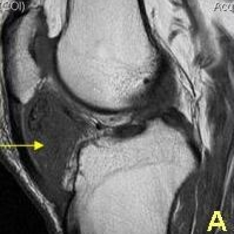
T1-weighted SE MR images show a mass with mixed signal intensity within the infrapatellar fat pad (Figs. 2 and 3).
T2-weighted SE MR image reveals high signal intensity within the mass that represents oedema and chondroid matrix (Fig. 4).
T2* MR image shows high signal intensity with central areas of low signal intensity that represents calcifications (Fig. 5).
DP FATSAT SE MR images shows heterogeneous high signal intensity lesion (Figs. 6 and 7).
Intracapsular chondroma, also known as extraskeletal chondroma, intracapsular osteochondroma and soft-tissue osteochondromais, is a rare lesion that results from extrasynovial metaplasia in the capsule or adjacent connective tissues. It is a benign cartilaginous soft tissue that may have foci of calcification, fibrosis and metaplastic ossification within it. It is considered by some as an end-stage form of Hoffa disease, given its similar location within the infrapatellar fat pad and similar pathologic feature of ossifying cartilaginous metaplasia.
It is characteristically located inferior to the patella and can produce bone erosion.
On radiographs it appears as a calcified mass within the infrapatellar fat pad.
Computed tomography usually shows a oval shaped soft-tissue mass with variable amount of ossification.
MR imaging: T2 and T2* weighted MR images demonstrates a heterogeneous mass with high signal intensity representing chondroid matrix or oedema and areas of low signal intensity representing calcification or ossification.
The most common differential diagnoses for this tumour are primary synovial chondromatosis and chondrosarcoma.
The treatment of choice is surgical excision.
Intracapsular chondroma


Based on the X-ray and MRI images of the knee joint provided by the patient, a soft tissue mass is observed beneath the patella (i.e., near the patellar tendon and fat pad area). The plain radiograph (X-ray) reveals irregular calcifications within the lesion, located at the inferior margin of the patella and within the anterior fat pad of the knee, possibly accompanied by local soft tissue prominence. A CT scan (if available) could further confirm its calcified or ossified components, typically appearing as an oval or irregular soft tissue density containing ossifications or calcifications.
On MRI, especially on T2-weighted and T2*-weighted sequences, the interior of the lesion shows heterogeneous signals, characterized by mixed high and low intensities: high intensities may correspond to cartilaginous matrix or mild edema, while low intensities may indicate calcified or ossified deposits. The lesion boundary is relatively well-defined, partially demarcating it from surrounding structures. There may be slight compression or even superficial cortical erosion of the patella or femoral condyle.
Based on the clinical and imaging characteristics, the following diagnoses should be considered:
Taking into account the patient's age (44 years), symptoms (anterior knee pain for one year, restricted range of motion, and soft tissue swelling in the infrapatellar region), imaging findings (prominent cartilaginous calcification or ossification in the infrapatellar fat pad presenting as a localized mass with heterogeneous MRI signals), the most likely diagnosis is Intra-articular Chondroma (Extraskeletal Chondroma).
If clinical ambiguity persists, further histological biopsy or intraoperative frozen section analysis could be performed to determine benign or malignant nature.
1. Treatment Strategy
Surgical resection is the first-line treatment, generally performed arthroscopically or with an open procedure aiming to completely remove the lesion. Given potential calcified or ossified components, thorough debridement is essential to reduce recurrence. If the lesion is large or anatomically complex, the surgical approach might require more detailed planning, possibly involving a “windowing” procedure or a small-incision method.
2. Rehabilitation and Exercise Prescription
Postoperative rehabilitation can be guided by a gradual approach following the FITT-VP principle (Frequency, Intensity, Time, Type, Volume, Progression):
This report is based solely on the available data for reference and does not replace in-person consultations or professional medical advice. If you have any concerns or if your condition changes, please consult a specialist and undergo further examinations to determine the most appropriate diagnosis and treatment plan.
Intracapsular chondroma