Diffuse large B-cell lymphoma of the femur with extensive involvement of adjacent soft tissues


Clinical History
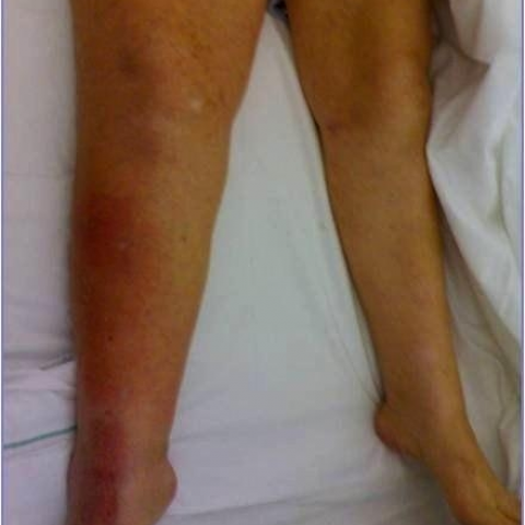
A 79-year-old woman, with diabetes mellitus and hypertension, came to our unit for complaints of right knee pain, extending towards the leg, which had been persistent for 5 months and unresponsive to analgesics. Physical examination detected right leg swelling with limited flexion-extension. Laboratory tests revealed lactate-dehydrogenase, fibrinogen and WBC count increments.
Imaging Findings
Ultrasound examination displayed: conspicuous right knee effusion involving superior recesses; synovial hypertrophy; hypoechoic mass in the middle-distal region of femur, displacing the middle, medial and lateral vast.
Femur X-ray showed: macular dishomogeneous structure of distal region, resulting from osteolytic lesions; cortical erosions of distal metaphyseal external surface.
CT scan, beside confirming radiographic findings, showed also: decalcification of medullary bone matrix; periosteal bone spicules protruding towards soft tissues; femur surrounded by muscular isodense masses with regular margins; vastus medialis swelling without densitometric alterations.
MRI, performed for suspected secondary lesion, displayed: diffuse femoral lesions (T1-hypointense/T2-hyperintense), invading periosseous soft tissues.
Following CT-guided biopsy, the diagnosis of diffuse large B-cell non-Hodgkin’s lymphoma (NHL) was made.
Whole-body CT staging revealed: multiple lung nodules (up to 4-cm diameter), compatible with metastases; several focal splenic hypodense lesions (from few mm to 2-cm diameter); involvement of mesenterial, right iliac, inguinal and femoral lymph nodes (up to 2.7-cm maximum diameter).
Discussion
Primary bone lymphoma is uncommon and affects mostly the metaphysis/diaphysis of long bones (usually femur). It occurs mainly in adult/elderly population, and its commonest histological phenotype consists of diffuse large B-cells. Clinical features include: localized or migrating pain and, less frequently, soft tissue swelling, palpable mass, pathological fractures and systemic “B” symptoms (Ann Arbor classification). In cases of disseminated disease, secondary bone involvement is difficult to distinguish from primary one, with radiological features being similar.
Radiological appearance of bone lymphoma is nonspecific and heterogeneous, making difficult the initial diagnosis. Usually, it appears as isolated or multiple radiolucent osteolytic lesions, often with sclerosis and poorly defined margins, characterized by permeative or moth-eaten pattern, corresponding to marrow and cortical replacement by lymphoma. Cortical destruction or erosion with soft tissue invasion is frequent and regarded as negative prognostic factor.
Bone scintigraphy appearances are also nonspecific, as lymphomatous lesions show a central area of decreased uptake, with increments at periphery.
CT is useful for: studying bone lesions; defining cortical destruction; evaluating soft tissue and marrow involvement; staging (evidence of local or distant dissemination).
MR allows local staging better than other techniques, particularly with regard to bone marrow and soft tissue involvement. MR signal intensity of bone lymphoma is dishomogeneous: isointense/hypointense to muscle on T1; hypo/iso/hyperintense to subcutaneous fat on T2; hypointense on T1/T2 for conspicuous fibrosis. Fast tumor growth with deficient vascular supply promotes necrotic areas, contributing to heterogeneity of signal intensity. Lymphomatous bone marrow is detected as a low signal on T1, due to fat replacement, in contrast with hyperintense normal marrow. Soft tissue involvement appears as isointense to muscle on T1 and hyperintense on T2, with diffuse enhancement. Short tau inversion-recovery (STIR) fat suppression sequences yield signal enhancement on T1 and T2.
PET scan is helpful in staging, and is superior to CT or MR in assessing remission.
Differential diagnosis includes: Ewing’s sarcoma, osteosarcoma, metastasis, osteomyelitis, multiple myeloma, and leukemic infiltrate. Only bone biopsy allows a correct diagnosis by immuno-histochemical analysis.
In the present case, the systemic dissemination of disease does not allow to discriminate between primary or secondary bone lymphoma. However, the extensive involvement of bone and adjacent tissues by lymphomatous lesion, together with the femoral location and histopathological diagnosis of diffuse large B-cell NHL, suggests a primary skeletal form. Whatever the origin, it is crucial for the radiologist to recognize lymphoma, despite its heterogeneous appearances, to allow timely and adequate therapeutic measures.
Differential Diagnosis List
Final Diagnosis
Diffuse large B-cell lymphoma of the femur.
Liscense
Figures
Ultrasound examination


Right femoral CT


Right femoral MR


CT-guided biopsy


Physical examination


Whole-body staging CT


Right femoral radiography

Medical Imaging Analysis Report
I. Imaging Findings
1. Bone destruction is observed in the right femoral shaft and near the knee joint. The lesion appears as an irregularly shaped lytic change, with some areas showing decreased density and unclear boundaries, along with signs of infiltration into the medullary cavity and soft tissues.
2. CT scans reveal erosion or destruction of the right femoral cortex. The lesion extends to the surrounding soft tissues, forming a soft tissue mass. Some areas appear “moth-eaten” or “diffuse infiltration” in nature.
3. MRI shows that the bone marrow signal is replaced by abnormal tissue: T1 often appears low or isointense, and T2 has mixed (low/medium/high) signal characteristics. Some fibrotic and necrotic areas are visible, leading to uneven signal distribution. Tumor infiltration in the soft tissue can also be seen, showing muscle-equivalent signal on T1 and slightly high signal on T2, with marked enhancement after contrast administration.
4. Ultrasound shows heterogenous echoes within the lesion, accompanied by surrounding soft tissue edema or exudation.
5. Further chest and abdominal CT suggests multiple enlarged lymph nodes and possible lesions or infiltrations in organs (such as the spleen), indicating multisite systemic involvement.
6. Laboratory tests show elevated lactate dehydrogenase (LDH), fibrinogen, and white blood cell counts, which correlates with high lymphoma activity.
II. Possible Diagnoses
- Primary Bone Lymphoma: This type of lesion commonly occurs in the metaphysis or diaphysis of long bones. In older patients, it is often diffuse large B-cell lymphoma. Imaging typically shows lytic or mixed bone destruction, with marrow and soft tissue involvement. Laboratory tests frequently reveal elevated LDH.
- Secondary Bone Lymphoma from Other Sources: If the patient already has a history of lymphoma or another organ lymphoma, the skeleton could be secondarily involved. However, imaging findings can be difficult to distinguish from primary bone lymphoma.
- Osteosarcoma or Other Malignant Bone Tumors: Conditions such as osteosarcoma or Ewing’s sarcoma can also show lytic or mixed bone destruction and soft tissue swelling, but clinical and pathological findings usually help differentiate these.
- Multiple Myeloma: Typically seen in older adults, often showing bone destruction. However, multiple myeloma commonly affects vertebrae, pelvis, and multiple sites in the limbs, with characteristic “M-spike” protein and other laboratory indicators.
- Infectious Osteomyelitis: Can also cause bone destruction and soft tissue swelling but is generally accompanied by obvious signs of inflammation (e.g., abscess, fever, significant neutrophil elevation in blood) and periosteal reactions on imaging.
III. Final Diagnosis
Given the patient’s older age, diffuse involvement of the right femur and surrounding soft tissue, the aggressive lytic destruction on imaging, along with elevated LDH and pathological immunohistochemical analyses confirming “Diffuse Large B-Cell Lymphoma (DLBCL),” the most likely diagnosis is:
Primary (or predominantly bone-involved) Diffuse Large B-Cell Lymphoma.
Although it is not absolutely possible to exclude secondary bone involvement based on imaging alone, the extensive bone and soft tissue lesions, pathological confirmation of DLBCL, and the clinical presentation all point towards primary bone lymphoma as the most likely diagnosis. Further precise staging and confirmation may involve whole-body PET-CT, bone marrow biopsy, or biopsies of other lesions as needed.
IV. Treatment Plan and Rehabilitation
1. Treatment Plan
- Chemotherapy: An immunochemotherapy regimen containing rituximab (e.g., R-CHOP) is commonly used as first-line treatment and can effectively control and reduce bone lymphoma lesions.
- Radiotherapy: Can be combined as a local treatment for areas with severe bone destruction or pain, helping reduce the risk of local recurrence.
- Surgical Intervention: If there is a pathological fracture or a significant risk of structural instability, surgical internal fixation, curettage of the lesion, or reconstruction may be considered to maintain limb function.
- Supportive Care: Manage comorbidities (e.g., diabetes, hypertension) by adjusting medications and providing symptomatic treatment. Closely monitor white blood cell counts, anemia indicators, and electrolytes.
2. Rehabilitation and Exercise Prescription (FITT-VP Principle)
During overall lymphoma treatment, rehabilitation and exercise programs should be individualized, especially when the bones are involved. It is essential to balance limb loading with the risk of fracture, prioritizing safety. Suggested measures include:
- Frequency: Based on the patient’s condition and overall status, plan low-intensity exercises 3–4 times per week initially, gradually increasing to about 5 times per week.
- Intensity: Begin with low-intensity activities (e.g., seated or bedside active movements, light resistance exercises) and gradually increase within tolerable limits. Use RPE (Rate of Perceived Exertion) around 11–13 as a guideline.
- Time: Start at 15–20 minutes per session, progressively increasing to 30 minutes. The total can be divided into multiple segments to avoid prolonged weight-bearing.
- Type: Prioritize activities that do not significantly increase fracture risk, such as seated or supine exercises, or low-impact training under protective measures (e.g., passive or assisted lower limb joint movements, recumbent stationary bike).
- Progression: Adjust exercise intensity and the duration of weight-bearing based on ongoing bone healing and overall condition, under the evaluation of a physician or rehabilitation therapist. If surgery and fixation are performed, progress from partial to full weight-bearing exercises as healing permits.
- Volume & Individualization: Keep the total exercise duration, frequency, and intensity within tolerable ranges. Continuously monitor joint pain, local swelling, or systemic fatigue, and adjust the regimen as necessary.
V. Disclaimer
This report is based on the available imaging and clinical data and is provided for reference only. It cannot replace an in-person consultation or professional medical advice. Specific treatment plans should be developed in conjunction with comprehensive patient evaluation and specialist assessment. If there are any questions or changes in the patient’s condition, please seek medical attention promptly.
Human Doctor Final Diagnosis
Diffuse large B-cell lymphoma of the femur.