


A 27-year-old man presented at the orthopedic department with a 3-year history of severe limitation on his locomotor capacity and persistent pain at the anteromedial side of the right thigh. These symptoms had been diagnosed and treated as a psychosomatic disorder because of his known mental illness.
Digital radiography revealed the presence of a large endosteal bone neoformation. The patient underwent radiological investigations such as CT that confirmed the presence of a gross neoplastic lesion involving the middle third of the right femur proximal diaphysis, expanding the cortical bone that appears discontinuous in several places, associated with periosteal reaction, most evident at the front side. This lesion measured approximately 177x73x69 mm and showed an inhomogeneous enhancement after contrast injection. The angio-CT documented bilateral patency of leg arteries. Subsequently T1, T2-weighted MRI and fat-suppressing sequences were performed for further characterization of the lesion, which confirmed the morpho-structural alteration of the proximal right femoral diaphysis caused by the lesion apparently constituted of a productive hypocellular tissue. At PET scan the neoplasm demonstrated a high captation of FDG. At last bone biopsy was performed for histological characterization.
Vascular bone tumours represent less than 1% of all bone tumours and their classification seems to be still evolving [1]. It was first described by Weiss and Enzinger in 1982 and often mistaken for a high grade malignant tumour. Considering their histological appearance and their biological behaviour these tumours are classified as intermediate between benign haemangiomas and malignant angiosarcomas and as proposed by Wanger and Wald they constitute their own subgroup between low grade and high grade bone vascular malignancies [2,3]. So the term epithelioid haemangioendothelioma was suggested to be used to designate these biologically “borderline” neoplasms. EHE occurs in the calvarium, spine, femur, tibia and feet of adults during the second or third decade. Chromosome analysis and molecular cytogenetic investigations showed that EHE is characterised by complex genetic rearrangements of 11q and 12q [4].
Usually, epithelioid haemangioendotheliomas present with pain and swelling and if present in the spine, they can cause radicular symptoms or paraplegia. Radiographically the majority of EHE show lytic bone destruction with associated alteration of matrix mineralisation, endosteal erosion and cortical thinning [5]. The contrast enhancement is easily evidenced by the Computed Tomography which can be applied for further characterisation of the neoplasm such as local and systemic extension and for vascular supply assessment of the tumour. At MRI the EHE usually presents with intermediate signal intensity on T1-weighted images, high signal on T2-weighted and homogeneous contrast enhancement without the detection of the typically serpentine vascular structures that would help in the differential diagnosis. FDG PET scanning in EHE, shows bone marrow involvement and determines the extent of the disease [6]. Although, the information gained through the imaging techniques are useful to identify and to stage the lesion, they are not entirely specific and the confirmation of the diagnosis is made possible only by biopsy and pathologic examination. The epithelioid histological subtype of haemangioendothelioma has epithelial-like cells lining the vascular channels that are large and cuboidal and contain abundant eosinophylic cytoplasm. Immunohistochemical study is helpful in confirming the diagnosis by identifying the markers for vascular endothelial cell. The clinical course and prognosis depend on uni or multifocality, histological differentiation and cytologic atypia. The overall survival is 89% in unifocal disease and 50% in multifocal involvement [7]. Surgical excision is considered the best treatment in addition or not with post-operatory chemotherapy or previous embolisation of the lesion [8].
Epithelioid haemangioendothelioma


Based on the provided X-ray, CT, MRI, and PET-CT images, there is a clearly visible lesion invading almost the entire mid to near-total segment of the right femur. The findings are as follows:
The lesion is extensive, involving most of the medullary cavity of the right femur and extending into the local soft tissue. The boundaries are not well-defined, and the enhancement pattern suggests a vascular tumor.
Considering the patient’s age (27 years old), three years of chronic progressive pain and functional impairment, and the above imaging characteristics, the following diagnoses are included:
In this case, EHE exhibits the typical “vascular origin between benign and malignant” features, matching both the imaging findings and the patient’s clinical presentation.
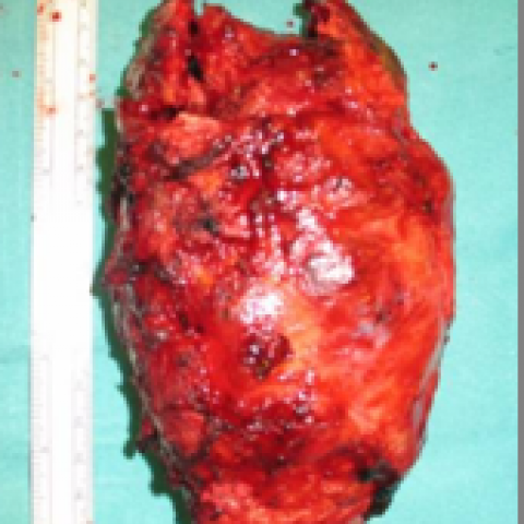
Based on the patient’s histopathological examination, the lesion is confirmed to be Epithelioid Hemangioendothelioma (EHE). Diagnostic clues include:
If there is a clinical suspicion but pathology is not definitive, further genetic testing or additional pathological analyses can be performed. In this case, pathology has definitively identified EHE.
The primary goal of the rehabilitation period is to maintain or restore limb function and reduce the risk of bone and soft tissue complications. The specific rehabilitation principles are as follows:
Following the FITT-VP principle (Frequency, Intensity, Time, Type, and Progression):
Throughout rehabilitation, closely monitor pain, surgical site stability, and local swelling. If any discomfort or signs of recurrence occur, seek medical attention promptly.
This report is based on the provided medical history and imaging data. It serves as a reference opinion and cannot substitute for in-person consultation or professional physician advice. Actual diagnosis and treatment should follow the comprehensive evaluation and recommendations of a specialist.
Epithelioid haemangioendothelioma