Surface osteosarcoma - case report


Clinical History
The patient was referred to our Hospital with a 6-month history of a painless swelling on the right thigh, associated with march disturbance. Upon physical examination, a firm mass was palpable on the anterior surface of the thigh. The patient denies traumatic events, weight loss or other associated signs/symptoms.
Imaging Findings
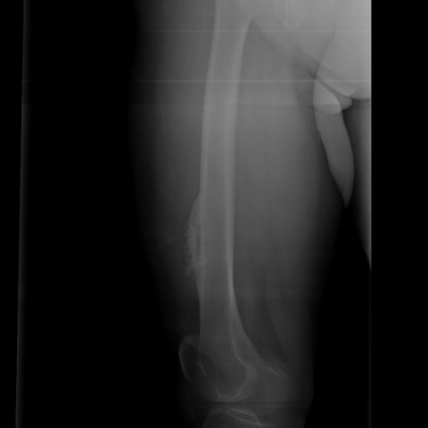
Antero-posterior and lateral plain radiography revealed an excentric lesion (7cm) on the distal antero-lateral femoral diaphysis, with spiculated periosteal reaction (fig.1).
On T1 –weighted MRI (fig.2) and FS T2–weighted MRI (fig.3) the distal antero-lateral femoral diaphysis showed a cortical lesion, measuring 6, 8x4, 6x2cm. This lesion presented peripheral high signal intensity on FS T2–weighted MRI. Central smaller foci of lower intensity were observed on both sequences. There was no intramedullary invasion. There were no skip lesions.
On CT scan (fig.4) there was calcified matrix extending into a low attenuation heterogeneous mass. There was an image suggestive of spiculated periosteal reaction. There was no evidence of intramedullary invasion.
The histological examination showed a low-grade neoplastic proliferation area, consistent with a superficial osteosarcoma (OS), according to clinical and radiological features. Histopathologic findings of surgical specimen (14 cm of distal femur) have confirmed to be a low-grade parosteal osteosarcoma.
Discussion
Surface OS constitute 4-10% of all osteosarcomas and have been subdivided in parosteal, periosteal and high grade surface OS.
Parosteal OS is the most common type of surface OS, accounting for 5% of all OS cases. It is seen in patients in their 3rd to 4th decades, with a female predominance. The tumor usually occurs in the metaphyses of long bones, characteristically affecting the posterior aspect of the distal femur. These lesions have an excellent prognosis. The classic appearance is a lobulated and exophytic mass adjacent to the bone, with central dense ossification, sharply delineated from the peripheral soft tissues, and cortical thickening. There is often a cleavage plane separating the tumor and adjacent normal cortex (string sign) that corresponds histologically to the periosteum interposed. The lesion arises on the outer fibrous layer of the periosteum, virtually never provoking a periosteal reaction. At MRI the ossified tumour is predominantly low in signal intensity on both T1- and T2-weighted images, similar to the appearance of the cortex. When the lesion is predominantly high in T2 signal intensity, the tumor is more likely to be of high grade.[1][2][3]
Periosteal OS has a peak incidence during the 2nd decade of life. These tumors show a strong predilection to arise in the diaphysis. These lesions are intermediate to high-grade and prognosis is usually worse than parosteal OS. Common radiological findings include a broad-based soft tissue mass, cortical thickening and extrinsic scalloping of the cortex, with periosteal reaction, often extended perpendicularly from the inner cortex. Hyperintensity on T2-weighted MRI and low attenuation on CT image are consistent with its cartilaginous component. In fact, histopathologically, the tumour is predominantly chondroid matrix with moderate cellularity, and reveals a large amount of cartilage matrix undergoing calcification, with areas of moderately differentiated osteoblastic osteosarcoma (osteoid formation). Invasion into the medullary canal is considered rare and needs to be distinguished from areas thought to represent reactive changes. This point is very important in directing the extent of surgical resection [1][2][3].
High-grade surface OS is rare and commonly involves metaphyses and diaphysis of long bones, especially femur. The prognosis is similar to that of conventional OS. High-grade surface OS presents dense ossification, bone destruction and periosteal reaction in the majority of cases. This tumour is often difficult to distinguish from periosteal OS. However, it usually surrounds a much higher percentage of bone circumference and is more likely to invade the medullary canal [1][2][3].
Differential Diagnosis List
Final Diagnosis
Parosteal Osteosarcoma
Liscense
Figures
Plain Radiography
T1-weighted MRI

FS T2-weighted MRI

CT scan

Medical Imaging and Clinical Analysis Report
1. Imaging Findings
Based on the provided X-ray, MRI, and CT images of the lower limb, a significant mass is observed in the anterior soft tissue area of the right thigh. The main characteristics are as follows:
- On the X-ray: A localized protrusion-like change on the bone surface is visible, with some lesions appearing as sclerotic densities and relatively clear boundaries. No obvious sign of intramedullary involvement is noted.
- On the MRI axial (T1 and T2-weighted) images: The main part of the lesion mostly shows low-to-medium signals, with some areas possibly showing high signal intensity, suggesting soft tissue or a small amount of edema. At the junction with the bone cortex, a locally thickened cortical layer or a thin boundary (“string sign”) can be seen.
- On the CT axial images: The lesion mainly presents as high-density ossification, with no significant expansion of the surrounding soft tissue component. No evident sign of medullary cavity invasion is observed so far.
2. Potential Diagnoses
Combined with the patient's medical history (62-year-old female, 6 months of painless swelling, no obvious history of trauma or weight loss) and the imaging characteristics, the following diagnoses are considered:
- Parosteal Osteosarcoma:
This is the most common type of surface osteosarcoma, frequently occurring in adults, often found on the distal femur (commonly posterior, but the anterior side can also be involved). The tumor often presents with an exophytic growth pattern and shows dense or lobulated ossification. A “string sign” often separates the normal bone cortex from the tumor. The patient's age and imaging findings are consistent with this condition. - Periosteal Osteosarcoma:
More often occurs in younger patients (around the second decade of life) and can be of moderate to high malignancy. It commonly shows marked periosteal reaction with a cartilaginous matrix and occurs in mid-diaphyseal regions. The presentation here does not fully match, but it remains a differential diagnosis. - High-grade Surface Osteosarcoma:
On imaging, there would typically be more bone destruction, a prominent periosteal reaction, and more extensive soft tissue infiltration. The prognosis is similar to that of conventional osteosarcoma. Although parts of this lesion show dense ossification, there is no overall indication of highly aggressive changes, so it is listed only as a differential diagnosis. - Chondrosarcoma or other cartilage-origin tumors:
These typically show “ring” or “arc-like” calcifications on imaging and should be considered in older patients. However, in this case, the main finding is the dense osseous lesion with exophytic growth from the surface, and the cartilaginous features are not prominent.
3. Final Diagnosis
In summary, taking into consideration the patient's age, sex, a gradually enlarging painless mass, and imaging findings of dense ossification with exophytic growth, along with a relatively clear boundary between the lesion and the cortex, the most likely diagnosis is:
Parosteal Osteosarcoma.
For a definitive diagnosis, a pathological biopsy is recommended to further clarify the histological grade and specific characteristics of the tumor.
4. Treatment Plan and Rehabilitation
Treatment Strategy:
- Surgical resection is the main treatment: For surface osteosarcomas, the resection should ensure a tumor-free margin. Whether to perform a wide excision or consider joint replacement depends on the extent of the lesion, including cortical and medullary involvement.
- Neoadjuvant or adjuvant therapy: If pathological grading indicates high-risk characteristics (e.g., high-grade malignancy), adjuvant chemotherapy may be considered. For low-grade lesions, surgical removal is often the primary approach.
- Individualized comprehensive treatment: Preoperative assessment of overall health status and any comorbidities should be performed, and the plan should be formulated in accordance with imaging and pathological findings.
Rehabilitation and Exercise Prescription:
A gradual rehabilitation program after treatment (especially surgery) is crucial. The following is recommended:
- Early Rehabilitation (0-6 weeks post-op):
- Focus on surgical wound healing and initial restoration of joint range of motion. Under the guidance of a physician or physical therapist, passive or assisted-active joint exercises can be performed.
- Depending on tolerance, conduct local isometric muscle contractions (avoiding large-range movements) to prevent muscle atrophy.
- Avoid weight-bearing or excessive range-of-motion activities. Keep the affected limb safe and prevent secondary injury.
- Mid-term Rehabilitation (6-12 weeks):
- Once bone healing and soft tissue repair are confirmed, gradually introduce functional exercises with posture control, such as sitting and standing training and light weight-bearing ambulation (assistive devices may be needed).
- Consider low-impact aerobic exercises, such as a stationary bike or an upper-body rowing machine. Training for overall cardiopulmonary function is particularly important.
- Strengthen core and trunk stability exercises, steadily improving strength and coordination.
- Late Rehabilitation (3 months to 1 year):
- Based on satisfactory bone and soft tissue recovery, gradually increase weight-bearing on the affected limb, e.g., short-distance walking and squats.
- Progressively increase the difficulty and frequency of strength and flexibility training, while closely monitoring for pain, swelling, or other issues, and adjust as needed.
- Set individualized exercise goals according to the patient’s baseline fitness and functional needs, avoiding excessive loads.
Guided by the FITT-VP principle (frequency, intensity, time, type, progression, and individualization), it is suggested to start with training three to four times per week at a tolerable or light-to-moderate intensity for about 20-30 minutes each time, and then gradually increase the duration and intensity as rehabilitation progresses. If the patient has reduced bone density or lower cardiopulmonary function, initial loads should be decreased, and close communication with the primary physician and rehabilitation specialist is essential. Periodic imaging follow-up is also recommended.
5. Disclaimer
This report provides a reference medical analysis and should not replace a face-to-face consultation or the advice of a professional medical institution. Specific treatment regimens should be formulated by the patient in consultation with specialist physicians, taking into account individual circumstances.
Human Doctor Final Diagnosis
Parosteal Osteosarcoma