A case of hibernoma


Clinical History
A 43-year-old female patient with lumbar spine fibrous dysplasia associated to iliopsoas incidentaloma.
Imaging Findings
A 43-year-old female patient presented to our department with low back pain which did not respond to painkillers and ambulation difficulties.
After close neurological examinations and L5 biopsy, diagnosis of fibrous dysplasia was made. She underwent surgical operation, then a PET was performed, showing active metabolism in the ilio-psoas muscle belly (Fig. 1).
CT of the lesion, which appeared as a hypointense mass, suggested a lipoma-like lesion (Fig. 2).
Then biopsy defined the nature of the lesion as a hibernoma.
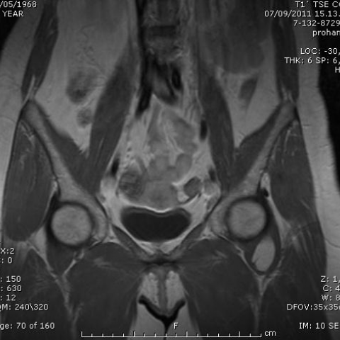
MRI was performed showing a well-defined mass, isointense to subcutaneous fat in FSE T1 images, high signal in T2 TSE images and no full suppression on fat-saturated T2-weighted images (Fig. 3).
Surgical treatment of the lesion is being planned.
Discussion
Hibernoma is a rare and asymptomatic benign tumour of muscles and soft tissues which arises mostly in adults from the remnants of fetal brown adipose tissue [1].
It usually occurs between ages 20 and 50 years.
In the adult, brown fat is usually found as persisting vestigial remnants along the oesophagus, trachea, posterior neck, interscapular area and around the great vessels of the mediastinum.
The most common anatomic locations include thigh, shoulder, back, neck, chest, arm and abdominal cavity/retroperitoneum; but they also occur in sites where brown fat is usually absent.
Hibernomas are slow-growing, painless neoplasms which do not recur. No malignant potential is known.
The hibernoma has been described as well encapsulated, tan-brown lobulated tumour. However, infiltration of adjacent structures, particularly striated muscle may be present [2-3].
Four morphologic variants of hibernoma were identified: typical; myxoid; spindle cell, and lipoma-like. The tumour is divided into lobules by thin septa. There are three cell types: large coarsely vacuolated cells, large finely vacuolated cells with eosinophilic granular cytoplasm and mature univacuolated adipocytes.
CT and angiography provided the most helpful information.
Hibernomas are fatty, solid and vascular. They appear clearly on CT as contrast-enhancing densities and, in this aspect, CT is superior to USG in localising the mass.
This hypervascularity also makes angiography an ideal tool for evaluation, but at the same time can mislead clinicians into suspecting a malignant process.
MRI findings usually demonstrate well-defined, heterogeneous mass, slightly or clearly hypo-intense to subcutaneous fat on T1-weighted spin-echo images, with prominent thin low signal bands throughout the tumour [2]. Lesions fail to fully suppress on STIR or fat-saturated T2-weighted images. Hibernomas characteristically demonstrate marked enhancement after administration of gadolinium [5].
Ultra-structural patterns analysis may be useful in differential diagnosis.
Hibernomas are circumscribed and with multiple small vacuoles cytoplasm, fat necrosis is not circumscribed and cytoplasm is foamy.
Lipoblastoma is typical in children and cells are immature lipoblasts, hibernoma is very rare in children and characterized by brown fat adult cells.
Prominent “chicken wire” vascular pattern and mitotic activity is distinctive of liposarcomas.
Cross-striated glycogen containing cells are typical in rabdomyoma, stromal chondroids elements are present in chondroid lipoma.
Hibernomas are considered benign. Sometimes they tend to enlarge in size causing compression of the adjacent structures. In these cases complete excision is the treatment of choice [4].
Differential Diagnosis List
Final Diagnosis
Hibernoma
Liscense
Figures
CT image

PET image


MR images


Medical Imaging Analysis Report
I. Imaging Findings
The patient’s CT and MRI images reveal a clearly demarcated soft tissue lesion in the pelvis and hip region, located near the iliopsoas/iliac muscle. On CT plain scan, this lesion overall appears as a relatively homogeneous soft tissue density, slightly higher than subcutaneous fat but lower than normal skeletal muscle, with a few high-density septa inside. On contrast-enhanced scanning, there is notable enhancement of the lesion, indicating a rich blood supply. On MRI plain T1-weighted sequences, the lesion signal is slightly lower than normal subcutaneous fat, while on T2-weighted and fat-suppression sequences (STIR or T2 FS), it is not fully suppressed, showing partly higher or mixed signal. Thin septa and vascular structures are visible internally. After contrast administration, the lesion shows pronounced enhancement. The margin of the lesion is relatively well-defined, yet there are signs of local extension or minimal infiltration into muscle fiber intervals.
Meanwhile, some fibrous changes in the lumbar vertebra are observed, consistent with the previously mentioned “fibrous developmental abnormality (fibrous dysplasia of bone)” findings. The changes appear benign, without obvious bone destruction. If no significant symptoms or progression of the lesion occur, continued follow-up observation is recommended.
II. Potential Diagnoses
- Hibernoma: Based on CT and MRI findings showing abundant blood supply, fatty signals that are not fully suppressed on fat-suppression sequences, and the typical age range (the patient is 43 years old), hibernoma is a strong consideration. It is generally benign, well-delineated, and richly vascularized.
- Lipoma or Atypical Lipoma/Well-Differentiated Liposarcoma: Lipomas typically have signals nearly identical to subcutaneous fat and might not present abundant vascularization. When numerous septations and notable enhancement are present, liposarcoma should be considered, although pathological confirmation is often required.
- Other Mesenchymal Tumors: Including myxoid lipoma, fibrolipoma, etc., which are less common and necessitate pathological examination for definitive diagnosis.
Due to the unique fatty component and rich blood supply observed on imaging—which closely aligns with the classic characteristics of hibernoma—hibernoma is the primary consideration.
III. Final Diagnosis
Considering the patient’s age, imaging features (abundant blood supply, partial fatty component not fully suppressed on fat-suppression sequences, relatively clear demarcation, internal thin septa, and strong enhancement), and the literature on hibernoma imaging, the most likely diagnosis is “Hibernoma.” If further confirmation is required, a biopsy or postoperative pathological examination may be considered.
IV. Treatment Plan and Rehabilitation
1. Treatment Strategy
- Follow-Up Observation: If the lesion is small with mild or no symptoms, periodic imaging evaluations can be conducted to monitor any progression.
- Surgical Resection: If the lesion enlarges, causing compression symptoms to adjacent structures (such as pain or restricted movement), or if there are suspicions of malignant transformation (though rare), complete surgical resection for both treatment and pathological clarification is advised.
- Postoperative Care: Hibernoma is benign, with a low recurrence rate, and complete excision typically achieves a cure.
2. Rehabilitation and Exercise Prescription
For patients who undergo surgical resection, postoperative rehabilitation is generally divided into the following stages:
- Early Stage (Postoperative Weeks 1–2): The emphasis is on protective recovery. Moderate passive and active lower limb movements can be performed, while avoiding large ranges of flexion-extension, excessive weight-bearing, or twisting motions. Light active flexion and extension of the hip and knee joints can be done in bed or from a seated position to maintain muscle tone.
- Intermediate Stage (Postoperative Weeks 2–6): In the absence of notable symptoms or complications, gradually increase walking distance and introduce mild resistance exercises, such as using resistance bands for isometric and isotonic training of the quadriceps, iliopsoas, and other relevant muscle groups. Movements should be performed slowly and progressed gently, with intensity adjusted based on pain and fatigue levels.
- Late Stage (Postoperative Weeks 6–12): If the incision site has healed well and no significant discomfort is reported, progressively transition to more comprehensive lower limb strengthening and core stability exercises, such as bridging and core muscle training. At this stage, moderate aerobic activities (e.g., brisk walking, elliptical trainer) may be introduced to aid in restoring cardiopulmonary function.
For patients not undergoing surgery (only under follow-up), if mild local discomfort arises, targeted muscle strengthening and stretching exercises can be considered, ensuring no undue trauma or excessive load, while maintaining proper lumbar and hip function.
Throughout the rehabilitation process, the “FITT-VP” principle should be followed, adjusting Frequency, Intensity, Time, Type, Volume, and Progression according to individual conditions. Each training session should be designed to avoid significant pain or fatigue, progressing step by step.
Disclaimer
This report is solely a reference-based medical analysis derived from existing imaging and clinical information. It should not be taken as a substitute for in-person medical evaluations or professional diagnoses and treatment recommendations from qualified physicians. Specific diagnosis and treatment plans must be based on thorough assessments of the patient’s detailed medical history, laboratory results, and pathological findings by specialist clinicians.
Human Doctor Final Diagnosis
Hibernoma