Carpometacarpal bossing: MRI findings


Clinical History
This 32-year-old female patient presented at our institution with bilateral swelling at hands dorsum, with bony consistence at palpation. The protuberance was located between II and III ray, at carpo-metacarpal level. Swelling became occasionally painful, often in association with hand overuse. A MR examination was performed.
Imaging Findings
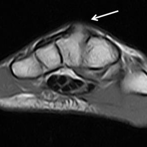
MR examination of both wrists, performed with a low-field (0.24 T) extremity-dedicated MRI system, shows signal alteration and a bony protuberance on the dorsal side between the base of the third metacarpal bone and the capitate, with aspect of neoarticulation: on sagittal images a bony bridge can be seen; alteration of the normal joint profile can be observed too (Fig. 1, 2). The tendons and the bone marrow seemed to return normal signal.
Discussion
Carpal boss is a rare disease, without gender predominance, characterised by the presence of a bony protuberance localised on the dorsum of the hand, in the carpal-metacarpal zone; it presents as a tough, painless swelling at physical examination.
The term “carpal boss” was coined by Fiolle in 1931: he firstly described this lesion as an exostosis at the base of II and III metacarpal bones. The origin of carpal boss is unknown and many theories were proposed during the years: the condition may represent a degenerative osteophytic process as well as the presence of an accessory bone.
In 1953, O’Rahilly described the presence of accessory ossicles in the carpal zone: one of this, the os styloideum, normally disappears during fetal development. The persistence of an accessory ossification centre can give rise to accessory bones like an “os styloideum”, located in the quadrangular trapezoid–capitate–metacarpal joint: it can interfere with the normal biomechanics of surrounding joints, leading to degenerative osteoarthritis, especially after repetitive stress [1].
The “os styloideum” may occur either as an isolated unit (articulated with one or more bones), or fused with one or more bones of the carpal or metacarpal zone; it has been proposed that this lesion could interfere with the normal biomechanics of adjacent joints, and lead to degenerative osteoarthritis, which can be symptomatic or not.
This bony prominence may dislocate extensor tendons and induce inflammation of these tendons and of surrounding soft-tissues [2].
Plain radiography, especially in lateral view, and computed tomography are fundamental modalities: they can visualise the bone protuberance, the joint profile and cortical erosions.
MR can provide a panoramic visualisation and highlight bony abnormalities that could lead to the formation of the bony bridge between the metacarpal base and the carpal bones and to subsequent complications: degenerative osteoarthritis can be visualised as an alteration of the normal joint profile on T1-weighted sequences, with an area of bone marrow oedema, hyperintense on T2-weighted and STIR sequences. MR can also show acute soft-tissues and extensor tendons inflammation [3].
Differential Diagnosis List
Final Diagnosis
Carpo-metacarpal bossing
Liscense
Figures
Left hand


Right hand


Medical Analysis Report
I. Imaging Findings
Based on the provided MRI sequences (axial and sagittal images, etc.), a noticeable bony protuberance is observed at the joint of the second and third metacarpals and adjacent carpal bones (such as the capitate and trapezoid). It appears as a lesion with low to intermediate signal intensity and fairly clear margins. Mild prominence or swelling is noted in the surrounding soft tissues, with a somewhat increased signal intensity suggestive of possible soft tissue inflammation or tenosynovitis. On T2-weighted and STIR sequences, a local mild hyperintense area is noted at the bone–cartilage interface, possibly related to mild osteoarthritic (degenerative) changes or bone marrow edema. Overall, no obvious fracture lines or large-scale soft tissue destruction are observed.
II. Potential Diagnoses
- Carpal Boss: This bony prominence typically forms at the base of the second and third metacarpals at the carpal–metacarpal joint, presenting as local swelling of a bony nature, with or without pain. Imaging often shows localized bony overgrowth, with or without mild joint degeneration.
- Degenerative Osteophyte (Osteoarthritic Osteophyte): Long-term mechanical friction or joint degeneration may result in osteophytes. On imaging, these can appear similar to Carpal Boss and require correlation with clinical symptoms and precise lesion location for differentiation.
- Ossified or Calcified Ganglion Cyst: A ganglion cyst usually manifests with soft tissue or fluid-like signals; however, the presence of calcification or ossification can produce high-density or abnormal signals on imaging and should be differentiated from a Carpal Boss.
III. Final Diagnosis
Combining the patient’s complaints (bilateral dorsal hand swellings, occasional pain, palpable bony texture without clear fluctuation) and imaging findings (localized bony prominence at the base of the second and third metacarpals near the radial or dorsal aspects of the carpal–metacarpal joint, with local soft tissue changes), the most likely diagnosis is Carpal Boss.
IV. Treatment Plan and Rehabilitation
1. Treatment Strategy
- Conservative Treatment:
- Use of wrist supports or elastic bandages to limit excessive wrist extension or friction at the protruding area, thereby reducing local irritation.
- Local physical therapy, warm compresses, and non-steroidal anti-inflammatory drugs (NSAIDs) can help relieve pain and inflammation.
- If there is notable inflammation, a local injection (corticosteroid + anesthetic) under sterile conditions may be considered to reduce pain and inflammation.
- Surgical Treatment:
- In cases of persistent or severe pain, recurrent inflammation, or functional impairment unresponsive to conservative treatment, surgical removal of the bony prominence or debridement of the affected area can be considered.
- Postoperatively, a cast or splint may be used for stabilization, with rehabilitation exercises introduced at an appropriate stage.
2. Rehabilitation/Exercise Prescription Suggestions
During both conservative management and postoperative rehabilitation, progressive functional exercises can be performed following the FITT-VP principles (Frequency, Intensity, Time, Type, Progression, Personalization):
- Frequency: Conduct hand and wrist functional exercises once or twice daily, gradually increasing to two or three times per day based on symptom improvement.
- Intensity: Start with low-intensity exercises, avoiding significant pain or discomfort. Light resistance training using grip balls or resistance bands can improve hand strength and wrist stability.
- Time: Each session should initially last about 5–10 minutes, gradually increasing to 15–20 minutes as tolerance improves.
- Type: Emphasize finger dexterity exercises (e.g., pinching, grasping, finger opposition), wrist range of motion (flexion, extension, pronation, supination), and stretching/relaxation exercises. Gentle grip ball exercises may be added if pain is not severe.
- Progression: Increase training load incrementally every 1–2 weeks, depending on symptoms and recovery status. If pain increases notably, reduce or pause training intensity and consult a physician or rehabilitation specialist.
- Personalization: Adjust the exercise schedule and program based on individual pain thresholds and recovery capacity. If other hand or systemic conditions are present, follow specialized advice from healthcare professionals or rehabilitation specialists.
Additionally, consider wearing braces or splints during exercises to stabilize the local joints and prevent secondary injury.
V. Disclaimer
This report offers a reference-based analysis derived from the provided medical history and imaging data and is not intended to replace an in-person consultation or professional medical advice. If you have further questions or your symptoms worsen, please seek prompt medical attention from a hand surgeon, orthopedic specialist, or other relevant healthcare provider, and follow professional guidance for subsequent treatment and rehabilitation.
Human Doctor Final Diagnosis
Carpo-metacarpal bossing