Transmural thoracic lipomatosis associated with congenital anomaly of the rib: A rare entity


Clinical History
A 50-year-old man was referred for evaluation of dyspnoea of 18 months’ duration. No history of smoking, but employed in a factory environment where dilute hydrochloric acid is used. Previously diagnosed with Obstructive Sleep Apnoea-Hypopnoea Syndrome (OSAHS), treated with Continuous Positive Airway Pressure (CPAP) with a favourable response.
Imaging Findings
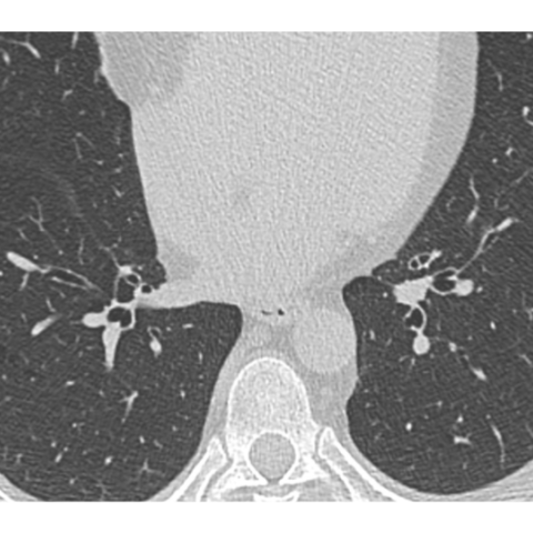
CT scan shows enlargement and sclerosis of the right seventh rib with a dysplastic aspect over its entire length with ossification almost to the sternocostal junction (Figure 1). Posteriorly, the rib shows slight periosteal irregularity and an image of small fragmented sessile exostosis or with adjacent heterotopic ossification (Figure 4).
Around the rib, there is a diffuse fatty infiltration of the thoracic wall, more prominent in the posterior half of the thorax (6th intercostal space) (Figures 3, 5a, 5b), extending medially and touching the pleura. There is also another area of lipoma, better defined anterolaterally, of lenticular morphology, in the fatty plane between the intercostal muscle and the oblique (Figure 6).
Sequences taken after contrast administration show no obvious enhancement of the lipomatous proliferation.
As no enhancing non-fatty tissue was detected, the associated rib anomaly and the absence of related symptoms, a diagnosis of transthoracic lipomatosis associated with congenital rib anomaly was made. A wait-and-see approach was decided with MRI examinations, which confirmed the stability of the lesion.
Discussion
Lipomatous lesions are a common group of mostly benign growths derived from adipose tissue [1]. They may present as solitary or multiple masses, deep or superficial. They are seldom seen in the thorax, although they can be found in many locations. Deep lipomas tend to be less circumscribed than subcutaneous lipomas, and their contours are usually determined by the spaces they occupy without infiltrating adjacent structures.
Deep thoracic lipomas can be divided into three groups according to their location:
- Intrathoracic: entirely intrapulmonary, subpleural or endobronchial.
- Cervicomediastinal: from the mediastinum to the neck.
- Transmural: they have an intrathoracic and an extrathoracic component connected by a fatty isthmus through the intercostal space or, exceptionally, through a sternal defect [2].
Exceptionally, they are associated with adjacent bone abnormalities (localised gigantism or malformation). Such cases have been reported, most of them more than 20 years ago [3,4].
The pathogenesis of the association between lipoma and congenital bone anomalies is unclear. The most widely accepted theory is that the nerve is initially irritated by an unknown cause, leading to abnormal trophic influences that alter the normal development of the soft tissues and bones corresponding to that dermatomal level, ultimately resulting in lipomatosis and dysostosis.
Clinically, they present as asymptomatic thoracic masses, preferably postero-lateral. Due to their slow growth, the symptoms are often due to the mass effect: cough, dyspnoea [5], cardiac dysfunction.
Chest x-ray shows a well-defined mass with intra- and extrathoracic components, with a homogeneous fat density and some metaplastic calcification.
Chest CT confirms the fat attenuation of the mass, together with changes in the morphology and size of the vertebra and the rib of the metameres, with irritative hyperostosis of the adjacent costal cortex.
MRI is useful in describing the extent of the lesion and its relationship to adjacent organs, as well as identifying the fatty nature of the lesion. The administration of intravenous contrast media provides information about the enhancing pattern: the less the lesion enhances, the more likely is to be of benign origin.
In all cases, deep lipomas must be differentiated from liposarcoma or other lipomatous malignancies, and the distinction often requires anatomopathological analysis.
The management strategy is not established. If asymptomatic, radiological follow-up may be sufficient, but some authors recommend biopsy or surgical excision due to the possibility of well-defined liposarcoma [6]. Even if they are histologically benign, surgery is sometimes necessary if there are symptoms due to their size or location.
All patient data have been completely anonymised throughout the entire manuscript and related files.
Differential Diagnosis List
Final Diagnosis
Multifocal transmural thoracic lipomatosis
Liscense
This work is licensed under a Creative Commons Attribution-NonCommercial-ShareAlike 4.0 International License.
Figures


Medical Imaging Analysis Report
I. Imaging Findings
Based on the provided 3D CT reconstructions and transverse sections (including CT and MRI sequences), a fat-density or fat-signal mass can be observed in the posterolateral thoracic cavity. Part of its extent lies within the thoracic cage and may extend through the intercostal space into the lateral chest wall. Specifically, the following findings are noted:
- On CT, the mass appears as a low-density lesion with an attenuation value close to that of fat (approximately -100 HU), and relatively well-defined margins.
- Local shape or size changes are noted in the ribs and adjacent vertebral body, with suspicious “irritative hypertrophy” of the rib cortex on imaging.
- MRI shows the lesion as high signal intensity on T1-weighted images and high signal on T2-weighted images. In some sequences, the signal is homogeneous and consistent with fat. After contrast enhancement, mild enhancement is observed around or within a small portion of the internal soft tissue, but there is no obvious enhancement in most of the lesion.
- No apparent invasive changes are noted in the adjacent lung tissue or mediastinal structures. The lesion overall demonstrates an expansive growth pattern.
II. Potential Diagnoses
Considering the patient’s symptoms (dyspnea lasting 18 months) and the fat-density lesion evident on imaging, along with previous clinical data, the following differential diagnoses are possible:
- Deep Lipoma: Commonly found in subcutaneous or deep muscle layers, potentially involving the chest wall or thoracic cavity and can be adjacent to bone. Characteristically shows fat-like imaging density/signal with relatively clear borders.
- Liposarcoma: Although less common, the malignant potential of fatty tumors must be considered. On imaging, liposarcomas may exhibit heterogeneous density or enhancement, with possible solid components, septations, or necrotic areas.
- Other Fat-Containing Lesions: Such as lipomatosis or lipomatous hamartoma, typically exhibiting fatty intensity/signal on imaging. Clinical and pathological correlation is necessary for definitive diagnosis.
Because the tumor is located between the chest wall and the thoracic cavity, with changes in adjacent ribs or vertebral bodies, it is prudent to exclude secondary bone lesions or other soft tissue tumors affecting the ribs during the diagnostic process.
III. Final Diagnosis
Considering the patient’s age (50 years), clinical presentation (chronic dyspnea over 18 months, no smoking history, previously diagnosed and controlled OSAHS), and imaging findings indicating a fat-containing space-occupying lesion, the most likely diagnosis is:
Deep Lipoma of the Chest Wall/Thoracic Cavity (Transmural Lipoma)
To definitively rule out low-grade liposarcoma or other specialized fatty tumors, a tissue biopsy or intraoperative frozen section analysis with final pathological diagnosis may be considered clinically.
IV. Treatment Plan and Rehabilitation Program
1. Treatment Strategies
- Conservative Observation: For patients without significant symptoms or only mild symptoms, and if imaging follow-up reveals no marked changes in the lesion, routine follow-up (e.g., every 6–12 months) may be considered.
- Surgical Treatment: If the lesion is large and causes pronounced dyspnea, skeletal deformation, or if there is suspicion of malignancy (e.g., liposarcoma), surgical resection may be warranted. Preoperative evaluation of cardiopulmonary function is recommended to minimize risk.
- Histological Examination: For any suspicious lesion, a needle biopsy or intraoperative frozen section can be done to rule out malignancy.
2. Rehabilitation/Exercise Prescription
Given that the patient has respiratory issues (previous OSAHS and current dyspnea related to chest wall changes), a properly designed exercise regimen during recovery should follow these principles:
-
Gradual Progression (FITT-VP Principle):
- Frequency: Exercise 3–5 times a week, increasing gradually according to tolerance.
- Intensity: Start with low-to-moderate intensity aerobic activities, such as walking or slow cycling, aiming for comfort and avoiding excessive fatigue.
- Time: Begin with 10–15 minutes per session. Increase by 5 minutes every 2 weeks, gradually reaching 30–45 minutes.
- Type: Primarily aerobic exercise with simple breathing exercises and stretching. Post-surgery, commence with basic breathing training (e.g., pursed-lip breathing, diaphragmatic breathing).
- Volume: Assess weekly total exercise time and intensity to make suitable adjustments as the body adapts.
- Progression: Safely raise goals based on improvements in dyspnea, gradually increasing exercise intensity.
- Individualization: If significant skeletal alterations exist, assess thoracic mobility thoroughly, include chest expansion exercises, and avoid large twisting movements or heavy lifting to prevent injury.
- Monitoring and Safety: Monitor heart rate, perceived exertion, blood pressure, and subjective symptoms during exercise. If chest tightness, shortness of breath, or abnormal tachycardia occurs, cease activity and seek medical attention.
V. Disclaimer
This report is based solely on the available imaging and clinical data and is intended for reference only. It does not replace in-person diagnosis or professional medical advice. Please consult a specialist or visit a hospital for a comprehensive evaluation and personalized treatment plan.
Human Doctor Final Diagnosis
Multifocal transmural thoracic lipomatosis