


A 78-year-old woman on clopidogrel was admitted with right groin and abdominal pain after suffering a fall. Whilst in X-ray she became haemodynamically unstable and her abdomen distended. Her blood pressure fell to 70/44 and her haemoglobin dropped to 5.0g/dl from 11.1 g/dl. She was resuscitated and taken to CT.
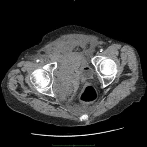
Pelvic and right hip images were ordered to ascertain any bony injury [Fig. 1]. Superior and inferior pubic rami fractures were identified. Chest and abdominal images were also requested but the patient became haemodynamically unstable whilst in the X-ray department and required fluid resuscitation. Once stable she was taken for CT thorax, abdomen and pelvis. CT imaging demonstrated fracture in both the superior and inferior pubic rami [Fig. 2]. Amorphous material was seen in the lower abdomen consistent with haematoma [Fig. 3a, b]. Free fluid can be seen to extend and surround the liver and spleen [Fig. 3c]. There was no aneurysm and no sign of vascular blush. There was no visceral injury but sign of right concomitant rectus sheath haematoma [Fig. 4].
Extraperitoneal bleeding has been attributed to pubic rami fractures with an increased risk associated with anticoagulation therapy [1]. Four cases of stable pubic rami fracture resulting in extraperitoneal haemorrhage have been reported in the literature [1-4]. There are no reports in the literature of pubic rami fractures causing a rectus haematoma.
Isolated pubic rami fractures are considered a stable pelvic injury. They are a common presentation in osteoporotic elderly female patients with an incidence of 25.6 per 100000 [1, 5]. Women are 4.2 times more likely to suffer this injury than men with a 5-year survival rate of 45.6% [5].
This case highlights an important complication of these seemingly benign fractures - haemorrhage. Stable fracture, according to the Tile Classification of pelvic fractures, is an intact sacroiliac complex. These fractures are not expected to bleed significantly. Common themes are illustrated between our case and prior cases. Firstly, the most likely source of bleeding is the inferior epigastric artery. Secondly, anticoagulation can significantly exacerbate the blood loss associated with so-called stable pubic rami fractures.
The anatomy of the region and the literature suggests that the most likely cause of haemorrhage is related to the pubic branch of the inferior epigastric artery [1-4]. Rectus haematomata develop as a result of bleeding into the rectus sheath due to direct tearing of the rectus muscle or from damage to the inferior or superior epigastric arteries. The posterior position make clinical diagnoses difficult as forming haematoma are difficult to palpate. The pubic branch of the inferior epigastric artery runs along inguinal ligament and the internal surface of the pubis before anastomosing with the obturator arterial system [3]. This branch has been described as the “Corona Mortis” due to difficulties in achieving haemostasis in this area [3, 6]. Cadaveric studies reveal significant variability in its anastomoses with 34% having an arterial connection, 70% having a venous connection and 20% showing a mixed picture [3, 6].
Anticoagulation is a known risk factor for rectus sheath haematoma [7] and can exacerbate bleeding from the inferior epigastric artery [1, 2]. Clopidogrel is an irreversible inhibitor of platelet aggregation [8], meaning uncontrollable bleeding associated with pubic rami fractures may require radiologically guided embolisation [1, 2, 9].
Increasing incidence of pubic rami fracture use of clopidogrel makes it increasingly important to remain vigilant over patients who have sustained this common injury. Signs of haemodynamic instability should be investigated appropriately for extraperitoneal haemorrhage and treatment should be directed at fluid resuscitation and/or embolisation as required.
Right superior/inferior pubic rami fracture; Massive extraperitoneal haemorrhage;Right rectus haematoma.


1. From the anteroposterior pelvic X-ray and pelvic CT scan, the integrity of the superior and inferior pubic rami appears compromised, suggesting fractures of the pubic rami.
2. Local soft tissue swelling and increased density are seen around the fracture, and a focal high-density shadow in the pelvic cavity indicates the formation of a local hematoma.
3. Further abdominopelvic CT scan reveals possible bleeding/hematoma in the lower abdominal wall area (near the pubic branch) and in the rectus sheath region, with partial signs of compression.
4. No obvious signs of parenchymal injury to solid organs (such as the liver and spleen) are observed, while the retroperitoneal space shows indications of localized bleeding.
1. Stable-type pubic ramus fracture with local soft tissue or rectus sheath hematoma:
Based on the fracture location (superior/inferior pubic ramus) and the vascular distribution of the lower abdominal wall, combined with the clinical use of antiplatelet medication (clopidogrel), the risk of bleeding is increased, presenting as an abdominal wall or pelvic cavity hematoma.
2. Other stable pelvic fractures with hemorrhage:
If the fracture line involves other adjacent pelvic structures connected to the pubic rami, there remains a possibility of local or intermittent bleeding. Attention should be paid to exclude injuries to the iliopubic or ilioischial ramus, etc.
3. Inferior epigastric artery injury or “Corona Mortis” hemorrhage (the pubic ramus may be a potential variant route):
Once this variant artery or its branches rupture, it can easily lead to uncontrolled bleeding. This needs to be differentiated on imaging.
Considering the patient’s age (78 years, female), the use of clopidogrel affecting coagulation function, and the current imaging findings of pubic ramus fracture accompanied by a significant hematoma, the most likely diagnosis is:
“Stable-type pubic ramus fracture with injury to the inferior epigastric artery or its branches, leading to hemorrhage within the rectus sheath (or extrapelvic).”
If necessary, further angiography or interventional examination can be performed to identify the bleeding vessel and achieve hemostasis via embolization.
1. Acute Phase Management:
• Enhance monitoring of vital signs, actively replenish fluids, correct anemia, and provide blood transfusion if needed.
• For suspected arterial bleeding, angiography and interventional embolization can be performed for hemostasis.
• Due to the patient’s use of clopidogrel, carefully assess the risks and benefits of continuing or temporarily stopping antiplatelet therapy.
2. Fracture and Pain Management:
• Generally, stable pubic ramus fractures can be managed conservatively, including immobilization, supportive rehabilitation exercises, and pain medication.
• Closely monitor bleeding in the pelvic area and the evolution of any hematoma, with repeat imaging if necessary.
3. Rehabilitation / Exercise Prescription:
• Initial Phase (Frequency, Intensity, Time, Type):
– Frequency: 1–2 times per day, short durations (10–15 minutes each session).
– Intensity: Mild active lower-limb movements, avoiding excessive weight bearing or intense exercise.
– Type: Static pelvic lifts, ankle pump exercises, and knee flexion/extension to prevent venous thrombosis and maintain basic joint mobility.
• Advanced Phase (Volume, Progression):
– As pain subsides and the hematoma resolves, gradually increase session duration to 20–30 minutes, 2–3 times a day.
– Moderately increase weight-bearing exercises for the lower limbs, such as short indoor walks with a walker or repeated sit-to-stand exercises.
– Closely watch for any renewed bleeding or worsening pain. Elderly patients are especially prone to fragile bone structure and limited cardiopulmonary reserve, so exercise intensity and form must be individualized.
Important Notes:
• For older patients on clopidogrel, take measures against falls during exercise or rehabilitation activities.
• Monitor blood pressure, heart rate, and hemoglobin levels to detect any recurrent bleeding or worsening anemia as early as possible.
Disclaimer: This report is based on the current imaging and clinical data as a reference and cannot replace an in-person consultation or professional medical advice. Diagnosis and treatment plans must be determined by a clinical physician based on the patient’s specific condition.
Right superior/inferior pubic rami fracture; Massive extraperitoneal haemorrhage;Right rectus haematoma.