Polyostotic fibrous dysplasia with facial bones involvement


Clinical History
10-year-old patient with previous known polyostotic fibrous dysplasia.
Imaging Findings
Multiple limb X-rays (Fig. 1-3) demonstrate the typical findings of polyostic fibrous dysplasia:
Expansion bone with typical "ground glass" (visualised in femur, tibia, radius and distal part of humerus) with slight bone deformity more appreciated on the tibia and femur.
Cortical thickening (metacarpals and phalanges - Fig. 4) and radiolucent medulla
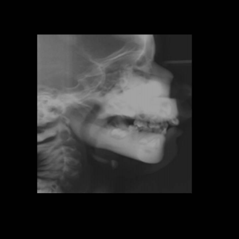
In X-ray and CT enlargement of facial bones with increased density (Fig. 5-7) can contribute to aesthetic changes and eventually the development of what some authors call lion face ("leontiasis faciei")
These changes are best demonstrated with three-dimensional CT images. (see Fig. 7 and 8)
Discussion
Fibrous dysplasia of bone is a genetic, non-inheritable disease, the normal bone and bone marrow is replaced with abnormal benign intramedullary fibro-osseous tissue, and can involve any bone in the body [1]
It can present in a monostotic or polyostotic form. Primarily affecting adolescents and young adults, it accounts for 7% of benign bone tumours [2]
Many of the asymptomatic lesions are found incidentally; the remainder present with symptoms of swelling, deformity, or pain [2].
The bone lesions may be associated with endocrine dysfunctions and café-au-lait spots, which is known as McCune-Albright syndrome. [3] Some complications, such as nerve compression and malignant transformation, are uncommon. [4]
The craniofacial involvement can pose a very difficult therapeutic problem due to localisation and uncontrolled proliferation followed by compression, both resulting in facial asymmetry, pain, cranial nerve deficiencies, alterations in hearing and loss of vision. [5]
Radiographic features of fibrous dysplasia of the skull and facial bones are described as three patterns: [6]
The pagetoid, or ground-glass, pattern is most common and consists of a mixture of dense and radiolucent areas of fibrosis. It also can be constituted by sclerotic homogeneous lesions whereas the cystic variety is characterised by a spherical or ovoid lucency surrounded by a dense bony shell. [6]
The most common appearance of fibrous dysplasia on CT is an expanded bone showing a ground-glass appearance and is usually straightforward [7]
MRI is not particularly useful in differentiating fibrous dysplasia from other entities as there is marked variability in the appearance of the bone lesions, and they can often resemble tumour or more aggressive lesions [7].
The MRI characteristics show signal intensity that is intermediate to low on T1-weighted images, intermediate to high on T2-weighted images, and shows heterogeneous enhancement after administration of gadolinium [8].
Diagnosis of fibrous dysplasia is important to recognise especially in the facial skeleton because of the possible unfavourable influence of the disease on stomatognathic system, laryngological disease, ophthalmic and neurological disease. [9]
Differential Diagnosis List
Final Diagnosis
Polyostotic fibrous dysplasia with facial bones involvement
Liscense
Figures
Polyostotic fibrous dysplasia

Polyostotic fibrous dysplasia- Hands X-ray

Polyostotic fibrous dysplasia- Lower legs X-ray

Polyostotic fibrous dysplasia - Lateral face X-ray
Polyostotic fibrous dysplasia- Coronal Face CT

Polyostotic fibrous dysplasia - Face CT 3D

Polyostotic fibrous dysplasia - Face CT 3D

Medical Imaging Analysis Report
I. Radiological Findings
Based on the provided X-ray and CT scans from multiple sites, the key observations are as follows:
- Long bones (e.g., femur, tibia, as well as some phalanges of the hand) exhibit irregular bone density with a “ground-glass” appearance in certain regions. There is also observable expansion of the bone and thinning of the local cortical bone.
- CT scans of the skull and facial bones show partial thickening of bone tissue with uneven density, manifesting various degrees of “ground-glass” changes and mild facial asymmetry.
- The maxilla and cranial base display interwoven areas of low and high density. The boundaries appear relatively defined, but the bone surface shows irregular protrusions, suggesting expansion and deformation.
- No definitive signs of fracture are noted, and there is no obvious soft tissue mass visible.
II. Potential Diagnoses
Based on the imaging findings and the patient’s previously confirmed history of polyostotic fibrous dysplasia, possible diagnoses or differential diagnoses include:
- Fibrous Dysplasia (Fibrous proliferative lesion): Highly consistent with the patient’s current presentation. This benign fibrous proliferative bone disorder commonly shows “ground-glass” appearances, especially in long bones and the craniofacial skeleton.
- Bone Fibroma or Osteoma (Other Benign Bone Tumors): Occasionally, these may appear similar to fibrous dysplasia on imaging. However, lesions tend to be more localized, and the confirmed multi-bone involvement in this patient more strongly supports fibrous dysplasia.
- Paget’s Disease (Late Stage): More prevalent in older patients, often accompanied by elevated serum alkaline phosphatase levels, which do not align perfectly with this patient’s age and presentation.
III. Final Diagnosis
Considering the patient is 10 years old, female, with a known history of “polyostotic fibrous dysplasia,” as well as the current imaging findings of multiple “ground-glass” lesions and bone expansion/deformity, the most likely diagnosis is:
Polyostotic Fibrous Dysplasia.
If there is any clinical concern about a malignant process or the activity level of the lesions, follow-up imaging or a biopsy may be performed to confirm the pathology.
IV. Treatment Plan and Rehabilitation
Treatment for polyostotic fibrous dysplasia should be tailored according to the extent of the lesions, severity of symptoms, and degree of functional impairment.
- Conservative Management: For stable lesions with no severe deformity or neurological symptoms, periodic follow-up and monitoring of bone changes may suffice.
- Surgical Intervention: In cases of significant bone deformity, functional impairment, or high risk of fracture, procedures such as corrective osteotomy or lesion curettage with bone grafting may be considered. Internal fixation can also be utilized to restore limb function and stability. For craniofacial involvement with nerve compression or significant functional impact, consultation with craniofacial or maxillofacial surgery teams is necessary to assess surgical indications.
- Pharmacotherapy: Bisphosphonates may help relieve pain or reduce high bone turnover in some fibrous dysplasia cases, though their ability to reverse established deformities is limited.
Rehabilitation/Exercise Prescription Suggestions (FITT-VP Principle)
- Frequency: Engage in low-impact aerobic exercises 3-5 times per week, such as swimming or stationary cycling.
- Intensity: Given that a child’s bones are still developing and bone integrity may be compromised by the disease, start with low to moderate intensity (e.g., RPE scale 11-13). Avoid high-impact or heavy-load activities.
- Time: Begin with 20-30 minutes per session, gradually extending to around 40 minutes, adjusting based on tolerance and recovery.
- Type: Focus on safety and protection of joints and bones; consider swimming, stationary cycling, light to moderate yoga, and core stability training.
- Progression: Once initial tolerance is achieved, gradually increase intensity or duration based on individual tolerance to maintain bone and muscle function, while minimizing excessive impact to affected bones.
- Volume & Pattern: Sessions can be divided into segments of 10-15 minutes to reduce fatigue. Schedule regular follow-ups to assess bone status and adjust exercise type and intensity as needed.
Throughout this process, regular follow-up with orthopedic, rehabilitation, and pediatric specialists is crucial. If pain, worsening deformity, or neurological symptoms occur, prompt medical evaluation is advised.
Disclaimer: This report is generated by an intelligent system based on the current imaging and medical history, and is for reference only. It does not replace in-person consultation or professional medical advice. For further inquiries, please consult a qualified healthcare professional.
Human Doctor Final Diagnosis
Polyostotic fibrous dysplasia with facial bones involvement