MR findings of a perineal angiomyxolipoma with pelvic extension


Clinical History
A 56-year-old man presented with a two-year history of a painless slowly growing mass in the perineum. On physical examination, there was a soft and well-demarcated oval-shaped mass in the anterior perineal area. Past medical history and laboratory studies were unremarkable.
Imaging Findings
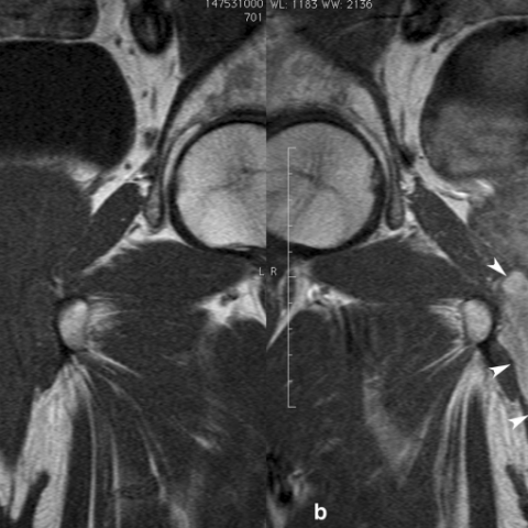
Pelvic MR imaging showed a well-defined perineal mass displacing the spongiosum corpus at the radix penis level, surrounding the spongiosum corpus at the pelvic diaphragm and extending to the pelvis surrounding the right prostatic apex (Fig. 1). The tumour showed intermediate signal intensity on T1-weighted images (Fig. 2), heterogeneous hyperintense signal on T2-weighted images (Fig. 1) and pronounced enhancement after intravenous gadolinium administration (Fig. 2). A swirled or layered appearance was seen on T2-weighted and gadolinium-enhanced images.
The patient underwent surgical resection of the perineal tumour. The mass was densely adherent to the spongiosum corpus and prostatic apex, and complete surgical resection was not possible.
The excised perineal specimen appeared well-defined with elastic consistency and light brown colour with whitish areas (Fig. 3a). Histopathologic examination was consistent with a vascular myxolipoma (angiomyxolipoma) (Fig. 3b and 4).
Discussion
Angiomyxolipoma (vascular myxolipoma) is a rare variant of lipoma, with very few cases reported in the literature in patients from 9 to 69 years [1, 2]. This tumour usually presents as a slowly growing, well-demarcated, painless subcutaneous mass on the scalp or extremities [2-5]. To our knowledge, our case is the first one presented as a large perineal mass extending from subcutaneous tissue to the pelvis through the pelvic diaphragm. This is a known route of tumoural extension between the pelvic cavity and the perineum [6], which is a challenge in management, given the proximity of the tumour to genitourinary and anorectal structures.
The histopathologic features of angiomyxolipoma include an admixture of a myxoid area with relatively few spindle-cells, mature adipose tissue without lipoblasts, and numerous thin- and thick-walled vessels [1-3]. Spindle cells in myxoid areas immunoreactivity for vimentin and CD34 [2, 3]. The mature adipocytes express S-100 protein, and the vessel walls expressed CD34 and SMA The tumour is negative for HMB45 [2, 3].
The MRI features of angiomyxolipoma have been previously described in only one case report, involving the suprapatellar area [4]. In this report, the tumour presents as a well-defined complex mass with a large peripheral fatty component and intense enhancement of the nonadipose central area. Our case appears to be quite different from previous reported cases, except for the well-defined margin and the intense enhacement after gadolinium administration. In our case, the mass showed intermediate signal intensity on T1-weighted images, although aggregates of microscopic fat within the tumour were seen on the pathological specimen. Microscopic fat would not be visible at MRI, unless chemical-shift imaging was performed. We also observed layered or swirled hypointense signal intensity within the hyperintense myxoid component of the tumour on T2-weighted MR images.
The swirled or layered appearance on MRI was attributed to the enhancement of the fibrovascular stroma. It usually develops in tumours that are stretched as they protrude through the pelvic diaphragm and has been reported as a characteristic finding in aggressive angiomyxoma and angiomyofibroblastoma [6, 7].
The tendency to infiltrate the surrounding soft tissue are key features in differentiating aggressive angiomyxoma from angiomyxolipoma [6]. Angiomyofibroblastoma was excluded in our case because it is miofibroblastic differentiation with HMB-45 inmunoreactivity, it is CD34 negative, the vessels are branching pattern and the tumour has epiteliod and plasmocitoid cells.
Differential Diagnosis List
Final Diagnosis
Angiomyxolipoma (vascular myxolipoma)
Liscense
Figures
Sagittal (a), coronal (b), and axial (c) TSE-T2-weighted

Coronal T1-weighted (a) and enhanced T1-weighted
Excised perineal specimen (a) and histopathologic examination (b)

Histopathologic examination

Medical Imaging Analysis Report
I. Imaging Findings
Based on the provided MRI data and clinical descriptions, a well-defined soft tissue mass is observed in the perineal region. Its specific characteristics are as follows:
- Location: The lesion is located in the anterior perineum, partially extending toward the pelvis and possibly passing upward through the pelvic diaphragm into the pelvic cavity.
- Shape: The mass is generally oval-shaped with heterogeneous internal signals. On T2-weighted images, high-signal areas with low-signal fiber bands or layered swirling patterns are seen, suggesting fibrous vascular septa.
- Signal Characteristics: On T1-weighted images, it shows intermediate signal, with minimal fatty signal not obviously apparent, although pathology confirms microscopic fat. On T2-weighted images, there is a prominent mucinous component with high signal, and after contrast enhancement, marked enhancement is observed, especially in the non-fatty areas.
- Adjacent Structures: The lesion occupies the deep soft tissue of the perineum, close to the urogenital tract and anorectal structures, but no evident signs of aggressive destruction are noted.
II. Possible Diagnoses
Considering the patient’s history (56-year-old male with a painless, slowly growing perineal soft tissue mass over the past two years) and the imaging findings, the following diagnoses or differential diagnoses can be considered:
- Lipoma (Conventional Type): Commonly seen in subcutaneous soft tissue, usually presenting a uniform fatty signal. However, in this case, the lesion contains more complex tissue components with abundant vascular and myxoid matrix, making a simple lipoma less likely.
- Aggressive Angiomyxoma: Often observed in the perineal and pelvic floor area, typically exhibiting ill-defined, infiltrative growth on MRI with spiral or layered vascular signals. However, such lesions generally demonstrate more pronounced local aggressiveness than seen here.
- Angiomyofibroblastoma: Commonly appears in the external genitalia and surrounding soft tissues, often featuring branching blood vessels. Immunohistochemistry may show HMB-45 positivity and atypical stromal cells, which do not match this case’s pathology results.
- Angiomyxolipoma: A relatively rare variant of lipoma characterized by myxoid stroma, mature adipose tissue, and an abundance of blood vessels. MRI typically reveals a prominent myxoid component coexisting with a small amount of fat and notable enhancement. In this case, pathological and immunohistochemical findings (CD34 positive, S-100 positive, HMB-45 negative, etc.) support this diagnosis.
III. Final Diagnosis
Considering the patient’s age, clinical history, imaging features, and immunohistochemical findings, the most likely diagnosis—in line with the pathological evidence—is:
Angiomyxolipoma.
IV. Treatment Plan and Rehabilitation Program
1. Treatment Strategy:
- Surgical Treatment: For larger or progressively enlarging angiomyxolipomas, complete surgical resection is recommended, aiming to remove the tumor capsule and any possible extensions to reduce the risk of recurrence. As the mass is close to the urogenital structures and rectum, meticulous protection of surrounding tissues is crucial during surgery.
- Postoperative Follow-up: After resection, regular follow-up using MRI or ultrasound is advised to monitor for local recurrence.
2. Rehabilitation and Exercise Prescription:
-
Early Postoperative Phase (Weeks 1-2):
- Incision Care and Basic Activity: Avoid excessive tension on the surgical site and keep the incision area clean and dry.
- Light Activity: Engage in short sessions of daily walking or low-intensity exercise, avoiding undue stress or pressure on the perineum.
-
Intermediate Postoperative Phase (Weeks 3-6):
- Gradual Lower Limb and Core Exercises: Activities such as light squats, leg raises, and simple functional training to improve muscle strength and stability.
- Pelvic Floor Exercises: Progressive Kegel exercises (e.g., contracting and relaxing the anal sphincter), gradually increasing the intensity and holding each contraction for a few seconds before releasing.
-
Late Postoperative Phase (Week 6 and Beyond):
- Aerobic Training: Gradually increase walking duration and speed according to cardiovascular fitness, or opt for low-impact exercises such as cycling.
- Continued Strength and Flexibility Training: If no contraindications exist, transition to moderate-intensity core and large muscle group workouts.
-
Application of the FITT-VP Principle:
- Frequency: 3-5 sessions per week, adjusted according to individual recovery.
- Intensity: Begin with low intensity, progressing gradually to moderate intensity.
- Time: Each exercise session should last 20-45 minutes, increasing gradually.
- Type: Initially focus on walking, pelvic floor exercises, and basic lower limb functional training; later introduce a variety of activities according to recovery progress.
- Progression: Increase the volume and difficulty of exercises steadily, guided by pain, tissue healing, and functional improvement.
- Precautions: Throughout rehabilitation, monitor for perineal pain, swelling, or other discomfort. If severe pain or signs of infection occur, seek medical evaluation promptly.
Disclaimer: This report is provided solely as a reference based on the current information available and does not replace in-person medical consultation or professional advice. If you have any questions or if your condition changes, please seek prompt medical attention for further evaluation and treatment.
Human Doctor Final Diagnosis
Angiomyxolipoma (vascular myxolipoma)