A case of a paraarticular chondroma in the Hoffa\'s fat pad


Clinical History
A 57-year-old man presented with a 3-month history of pain in his right knee. He reported also a lack of extension of the joint. He did not report a history of trauma. Physical examination showed a painful mass under the patella.
Imaging Findings
Radiograph in the lateral view shows a soft-tissue mass in the Hoffa’s fat pad; some central high density due to the calcification may be seen inside (Fig. 1).
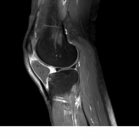
The subsequent MRI examination confirms the presence of a well-defined lesion in the infra-patellar fat pad. It presents intermediate signal intensity on sagittal T1-weighted images (Fig. 2).
On the axial fat saturation T2-weighted images the lesion shows heterogeneous, mainly high signal intensity (Fig. 3). On both T1 and T2 sequences the lesion presents some small areas of low signal intensity probably due to the calcifications.
After contrast administration a peripheral enhancement is seen (Fig. 4).
Discussion
Para-articular chondroma is a rare, benign soft tissue tumour [1, 2]. It arises from the capsule or the para-articular connective tissue of a joint. The pathogenesis of these tumours is still debated: it seems to be related to the cartilaginous metaplasia of the mesenchymal cells [1]. This lesion affects both men and women, between 10 and 80 years of age.
Although some cases have been described in the ankle, elbow and the hip joint, the knee is most frequently affected; this tumour is more frequently intra-capsular, involving the infra-patellar fat pad and only few cases have been described with an extra-capsular location [1, 2, 3]. Clinically, the tumour appears as a painful mass. The patient suffers from a lasting pain, with a time span from several months to many years, associated with a limitation of joint movement. On physical examination the lesion may be stable or mobile.
The plain radiographs may show a soft tissue mass; calcifications are visible in about 30-70% of cases. The pattern of calcification is mainly that of curvilinear and ring-like densities. Rarely, a peripheral rim ossification is seen. Secondary changes of the adjacent bone, such as cortical erosions and reactive sclerosis, have been reported in the literature.
On MRI the para-articular chondroma appears as a well-circumscribed mass isointense or hypointense relative to skeletal muscle on T1-weighted images. On T2-weighted images the tumour mainly appears hyperintense; on both T1 and T2 sequences the lesion may appear heterogeneous because of the central ossification that causes signal voids. After contrast injection the lesion exhibits more frequently a peripheral enhancement [3].
Para-articular chondromas must be differentiated mainly from other benign intra-articular and juxta-cortical calcified lesions, such as synovial chondromatosis [4] (in which lesions are multiple) periosteal chondroma (which shows severe cortical erosion) localized nodular synovitis and old haematomas. In differential diagnosis malignant lesions, such as synovial sarcoma and synovial chondrosarcoma, must be considered [1, 2].
The certain diagnosis must be confirmed by histopathologic examination.
Surgical excision is the treatment of choice, being careful not to injure the joint integrity. Despite the benign nature of the lesion, local recurrence has been reported; malignant transformation has never been referred. The small size of the lesion despite its long history, the nature of calcifications and the location itself, help to make the correct diagnosis in order to avoid whatever kind of unnecessary and aggressive surgical treatment.
Differential Diagnosis List
Final Diagnosis
Para-articular chondroma
Liscense
Figures
Lateral plain radiograph

Sagittal T1-weighted

Axial Fat Saturation T2-weighted

Sagittal Fat Saturation T1-weighted with gadolinium enhancement
Medical Analysis Report
I. Imaging Findings
Based on the provided knee X-ray and MRI images:
- X-ray: A distinct soft tissue mass is visible below the patella, with ring or arc-like calcifications in certain areas. No obvious bony destruction or severe cortical erosion is observed.
-
MRI:
- On T1-weighted images, the lesion usually appears isointense or hypointense, resembling the surrounding muscle signals.
- On T2-weighted images, the mass typically shows hyperintense signals; however, signal voids or mixed signals may be present due to internal calcification or local ossification.
- After contrast enhancement, the lesion predominantly demonstrates peripheral enhancement with relatively well-defined margins, indicating a likely benign lesion.
Overall, the lesion is located in the soft tissue around the joint (particularly beneath the patella, possibly involving the infrapatellar fat pad or surrounding tissues), with a relatively benign relationship to adjacent bone. Some degree of calcification or ossification is noted.
II. Potential Diagnoses
Taking into account the 57-year-old female patient with right knee pain for 3 months, limited range of motion, a palpable local mass, and the aforementioned imaging features, the primary considerations include:
- Para-articular Chondroma: A benign cartilaginous tumor often seen near the soft tissues around a joint. It may show ring or stippled calcification, typically appears hyperintense on T2-weighted images, and demonstrates peripheral enhancement after contrast.
- Synovial Chondromatosis: Characterized by multiple cartilaginous nodules and intra-articular loose bodies. However, this case shows a solitary or localized soft tissue mass, and the distribution pattern of calcification on X-ray differs from synovial chondromatosis.
- Periosteal Chondroma: Usually closely adheres to the bone cortex and shows marked cortical reaction, such as cortical thinning or erosion. This is not consistent with this case, which is primarily in the soft tissue with well-defined borders.
- Localized Nodular Synovitis and Chronic Hematoma: Both can present as soft tissue masses, yet they typically show fewer calcifications or cartilage-like signals.
- Malignant Possibility (e.g., Synovial Sarcoma or Chondrosarcoma): Although vigilance is necessary, the clearly defined margins, lack of aggressive features, and benign pattern of calcification make malignancy less likely.
III. Final Diagnosis
Based on the patient's age, symptoms, and imaging findings, the most likely diagnosis is: Para-articular Chondroma.
If clinical and imaging presentations remain uncertain, surgical resection and histopathological examination are recommended to confirm the diagnosis and rule out malignancy.
IV. Treatment Plan and Rehabilitation Program
- Surgical Treatment: Complete surgical excision of the lesion is the primary treatment of choice for patients diagnosed with or highly suspected of having a para-articular chondroma. Careful operative techniques are required to protect joint structures and avoid unnecessary damage to surrounding ligaments, tendons, and articular cartilage.
- Postoperative Rehabilitation:
- Early Phase (Post-op 1–2 weeks): Emphasize joint immobilization or limited activity. Include minimal passive joint mobility exercises under professional guidance (e.g., 10–20 degrees of flexion-extension) and isometric quadriceps exercises to maintain muscle strength.
- Intermediate Phase (Post-op 2–6 weeks): With good wound healing, gradually increase range of motion and muscle strengthening exercises, such as straight-leg raises, seated or supine flexion-extension training. Low-intensity cycling (with minimal resistance) may also be introduced to improve joint flexibility and muscle recovery.
- Late Phase (Post-op 6 weeks and beyond): Depending on functional recovery, progressively increase resistance training (e.g., exercises with resistance bands, weight-bearing squats) along with proprioception and balance exercises to enhance joint stability and overall lower-limb coordination.
- FITT-VP Principle:
- Frequency: Begin with 2–3 sessions per week, increasing to 3–5 times per week as tolerated.
- Intensity: Start with low resistance or low load, adjusting gradually according to joint pain and swelling.
- Type: Focus initially on range of motion exercises and isometric training; gradually incorporate resistance training and aerobic activities in later stages.
- Time: Each session starts at 20–30 minutes, eventually progressing to 40–60 minutes.
- Progression: Increase resistance or exercise duration incrementally based on individual response and recovery progress.
- Other Supportive Treatments: If postoperative knee pain or swelling occurs, consider physical therapies (e.g., cold compress for swelling relief, heat therapy to improve circulation) and short-term use of nonsteroidal anti-inflammatory drugs to control the inflammatory response.
Disclaimer
This report is a reference medical analysis based on the currently provided information and cannot replace in-person consultation or a qualified physician’s opinion. If you have any concerns or if your condition changes, please seek timely medical assistance for a comprehensive, personalized evaluation and treatment plan.
Human Doctor Final Diagnosis
Para-articular chondroma