Congenital vertebral segmentation abonormality in absence of other malformations


Clinical History
We present the case of a 31-year-old woman with chronic low back pain refractory to conservative therapies (analgesics and physical approaches) during the last six months. A pelvic misalignment and clinical signs of vertebral rotation were detected.
Imaging Findings
X-ray examination showed the presence of dorso-lumbar Congenital Vertebral Malformations (CVM) (Fig. 1).
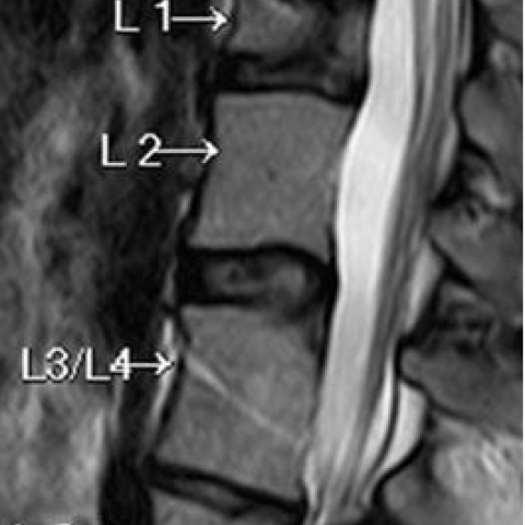
A T11-T12 disk bulging and vertebral segmentation and fusion abnormalities were well described with Magnetic Resonance Imaging (Fig. 2a). A L3-L4 vertebral block (Fig. 2a) and a L1 sagittal cleft ("butterfly vertebra") (Fig. 2b) were observed.
An anomalous origin of the 12th left rib from T11 was detected. In Fig. 3a a coronal image magnification shows the irregular morphology of T11 vertebra characterized by an antero-inferior wedge-shaped portion. An X-ray image magnification shows the anomalous origin of the left 12th rib from the T11 vertebra (Fig. 3b).
An abdominal ultrasound study excluded other visceral abnormalities.
Discussion
Vertebral segmentation refers to the embryonic developmental process, which will lead to the formation of the spine [1].
Different CVM classifications have been proposed, but no one has been able to include all phenotypes encountered in clinical practice (Table 1) [2, 3, 4, 5, 6, 7].
Genetic familiar transmission of diagnosed CVM has been documented in approximately 3% [8]. Chromosome abnormality or syndromes are estimated to be present in 30-60% of vertebral malformations [9].
In recent decades a series of gene alterations have been identified as responsible of mistakes in vertebral segmentation (Table 2) [10, 11, 12, 13, 14, 15].
In our case, after an accurate research in the literature, no similar phenotype has been described yet.
In fact in the same patient we described a segmentation defect (L1 "butterfly vertebra"), a fusion defect (L3-L4 vertebral block) and an anomalous origin of the 12th left rib from the T11 vertebra [16]. The last finding could be due to a fusion alteration of a T12 portion. The vertebral part, where the 12th left rib arises, is not completely fused with T11 vertebral body; furthermore we can't observe any left rib arising from the lower vertebral body. These observations lead us to think the 12th left rib arises from a vertebral particle which didn't fuse with T12 and migrated to the cranial vertebra.
CVM can be single or multiple and sometimes associated with known syndromes [16].
Isolated CVM may be asymptomatic or clinically appear with unexplained back pain or spine developmental abnormalities, after the skeletal maturation is completed [4, 6, 7, 8].
Sometimes CVM may cause a reduction of daily quality of life [17, 18, 19].
Therefore the early diagnosis is important with the aim to plan correct therapeutic strategies.
Imaging has a pivotal role. X-ray is the first imaging technique used to confirm the clinical diagnosis and to evaluate the seriousness of the pathologic condition [20].
Second level imaging is mandatory to well study the spine and organ systems.
Computed Tomography is recommended if CS is due to osseous abnormalities and for surgical planning [21]. MRI does not use ionizing radiations and is performed with increasing frequency because of the young age of the patients. MR multi-parametric study also allows detecting nervous system involvement [22].
Differential Diagnosis List
Final Diagnosis
Low back pain for congenital scoliosis caused by vertebral malformations.
Liscense
Figures
Antero-Posterior X-ray projection

The MRI examination allows to describe the vertebral malformations

The T11 morphological dysmorphism studied with MRI and radiographic examination


Table 1

Table 2

Medical Analysis Report
1. Imaging Findings
Based on the provided imaging (X-ray and MRI) and the patient’s clinical history, the following key features can be observed:
• In the lumbar spine series, the L1 vertebral body shows a “butterfly vertebra” change, suggesting a segmentation developmental anomaly.
• Vertebral fusion (block vertebra) is observed between the L3-L4 vertebral bodies, indicating a fusion defect.
• The 12th left rib originates from the T11 vertebral body, revealing rib development and vertebral accessory structure abnormalities; it is speculated that there may be a fusion anomaly at the corresponding T12 level.
• Mild pelvic asymmetry and signs of vertebral rotation are noted.
Overall imaging suggests multiple congenital vertebral malformations (CVM), with related variations particularly evident in the lumbar region and at the thoracolumbar junction.
2. Potential Diagnosis
Based on the above imaging findings and the patient’s clinical symptoms (chronic lower back pain for six months, with unsatisfactory response to conservative treatment), the following diagnoses or differential diagnoses can be considered:
(1) Multiple Congenital Vertebral Malformations: The presence of butterfly vertebra, block vertebra, and abnormal rib origin all suggest embryonic segmentation abnormalities consistent with congenital vertebral malformations.
(2) Spinal Dysplasia or Vertebral Malformations Related to a Specific Syndrome: Chromosomal abnormalities or certain genetic syndromes may present similar multi-system skeletal malformations. If the patient has other systemic abnormalities (not yet clarified), further investigation is warranted.
(3) Congenital Lumbar Transverse Process/Rib Anomaly or Hemivertebra Variants: Certain hemivertebra variants or transitional vertebra can be mistaken for similar findings; however, in this case, the presence of block vertebra, butterfly vertebra, and rib anomalies more strongly supports multiple congenital vertebral malformations.
3. Final Diagnosis
Combining the patient’s age, clinical symptoms, and the clear imaging findings (butterfly vertebra, block vertebra, and aberrant rib), the most likely diagnosis is:
Multiple Congenital Vertebral Malformations (CVM).
Currently, there is no evidence of other systemic or chromosomal abnormalities. Further examination is needed to rule out or confirm any associated syndromes or genetic factors.
4. Treatment Plan and Rehabilitation Strategy
Given that the patient’s symptoms significantly affect their quality of life and conservative treatment has been ineffective, the following treatment strategies should be evaluated comprehensively:
(1) Conservative Treatment
• Under specialist guidance, use analgesic drugs (e.g., non-steroidal anti-inflammatory drugs, NSAIDs) and muscle relaxants to alleviate pain and relieve muscle tension.
• Postural adjustment and brace support: If significant spinal instability or deformity causes pain, consider short-term bracing.
• Physical therapy: Emphasize spinal stability training and pelvic symmetry training. Manual therapy, traction, or soft tissue release can help relieve symptoms.
(2) Surgical Intervention
• If there are signs of nerve compression or progressive deformity caused by spinal instability, surgical correction or decompression may be considered upon medical evaluation.
• The surgical approach should be determined based on the degree of spinal rotation deformity, neurological involvement, and the patient’s overall health status.
(3) Rehabilitation and Exercise Prescription
Following the gradual progression (FITT-VP principle) and individualization, the recommendations include:
1) Frequency (F): Perform rehabilitation training 3-4 times a week, with adjustments based on fatigue and pain levels.
2) Intensity (I): Use low to moderate intensity exercises, avoiding heavy loads or violent twisting motions. Self-monitor pain and fatigue, and reduce or pause if significant pain occurs.
3) Time (T): Each session should last 20-30 minutes, including warm-up, core strengthening, flexibility exercises, and cool-down.
4) Type (T):
• Core stability exercises: such as plank and dead bug, ensuring the lumbar spine remains neutral.
• Joint range of motion and flexibility exercises: gentle stretching of the lumbar and pelvic muscle groups, avoiding excessive forward bending or twisting.
• Postural muscle strength training: gradually incorporate small equipment or exercise balls to enhance coordinated stability.
5) Progression (P): Gradually increase training time or difficulty based on pain relief and functional improvement, for example by increasing core training loads or extending stretching durations.
Safety Precautions:
• Avoid high-impact or intense rotational movements to prevent exacerbation of pain.
• If persistent worsening pain or lower limb numbness or weakness occurs during exercise, seek medical evaluation promptly.
Disclaimer
This report provides a reference medical analysis only and does not replace an in-person consultation or professional medical advice. The patient is advised to undergo further examinations and develop a treatment plan at a professional medical institution to achieve personalized and optimal clinical outcomes.
Human Doctor Final Diagnosis
Low back pain for congenital scoliosis caused by vertebral malformations.