


25-year-old pregnant, afebrile female with no significant background arrived due to back pain with no history of trauma.
The patient was send to the radiology department where an US and non-contrast MRI were performed. IV contrast and radiographs were not requested due to the patient's pregnany.
Ultrasonography showed hypoechoic lesion in the paravertebral musculature with small hyperechoic areas within, with acoustic shadow. There was significant peripheral hyperaemia on Doppler. (Fig 1)
On MR, the mass was well-defined, heterogeneous predominantly isointense to muscle on T1; hyperintense on T2 with hypointense foci. STIR showed surrounding oedema that extended into multiple soft tissue compartments. It also showed oedema in transverse process and hypointense peripheral area. (Fig.2-4)
The differential diagnosis included tumour and infection, therefore biopsy was performed. Biopsy did not show evidence of malignancy and serology and microbiology cultivation tests were negative as well.
After ten days, on follow-up ultrasound the lesion size was the same, hyperaemia had decreased and peripheral calcification became more evident. Due to the evolution myositis ossificans was our diagnosis. US follow-up demostrated increase of the peripheral calcification, that is the (zone phenomenon explained in 'Discussion'). (Fig. 5)
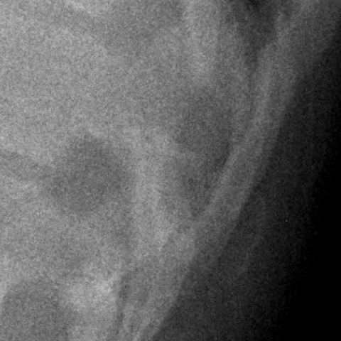
Later plain film showed the lesion as calcified on the periphery and not in the center.(Fig.6)
Myositis ossificans (MO) is a benign process characterised by heterotopic ossification usually with large muscles. Its importance stems from its ability to mimic more aggressive pathological processes, it may be clinically and histologically mistaken MO for a malignant soft tissue tumour[1] and this can lead to inappropiate management.
The aetiology and potential predisposing MO factors remain unclear. In some cases, non-causative factors can be identified, but in most cases it can occur as a result of muscular trauma, commonly in the thigh or arm.
There are several distinct stages of MO. In early stages histologically, the proliferation is composed of mesenchymal cells and fibroblasts, radiographs cannot show any anomaly, but in later stages, also called the maturation phase, bone production can be observed at the periphery of the lesion, radiographs may show the pathognomonic ossification surrounding a clear central area.This happens because the characteristic rim of calcification may be identified in 4-6 weeks after the injury.
This pattern of calcification is essential to establish the radiologic diagnosis, and allows differentiation from other mineralized lesions, such as sarcoma, that usually shows central calcification. If this peripheral calcification is not present, the diagnosis remains non-specific and an aggressive sarcoma or infection cannot be excluded.[2]
Computed tomography (CT) is more sensitive than radiography to detect calcification.
Magnetic resonance imaging (MR) appearance changes with the age of the lesion. We can differentiate between early and late stages[3]:
-Early lesions: peripheral mineralization is not visible or well-defined making the diagnosis more difficult. On T1, the lesion is isointense to muscle; on T2, signal intensity is high and secondaty to proliferating and cartilagenous components. Active lesions present enhancement after gadolinium injection and are surrounded by soft tissue oedema.
-Late lesions: both T1 and T2 show low signal intensity in the periphery corresponding to mature lamellar bone. The centre shows intermediate to high signal intensity. The difference between MO and sarcoma consists of the type of mineralization: whereas MO shows peripheral mineralization, sarcomas have a centre calcification.
Ultrasound is the most sensitive imaging modality to depict the zone phenomenon as it may suggest the diagnosis in early stage. There is a central, undifferentiated zone impossible to distinguish from sarcoma. This zone merges into oriented osteoid formation and finally into well-formed bone in the periphery of the lesion. The diagnosis has to be based not only on the history of the trauma, but also on other clinical and imaging outcomes.[4][5]
As MO is a benign process and there is no compelling evidence that malignant degeneration or tumour ever occurs, treatment is reserved for symptomatic lesions.
Myositis ossificans


The patient is a 25-year-old pregnant woman presenting with lower back pain, with no clear history of trauma. Considering her pregnancy, an ultrasound and a non-contrast MRI were performed. Ultrasound images show a relatively well-defined lesion located within the back muscles (paravertebral soft tissue), appearing roughly round or oval, with a fair margin. Ultrasound reveals partial calcification or strong echogenic rings at the periphery, forming a ring or “halo” sign around a relatively hypoechoic center. Doppler imaging indicates a small amount of blood flow around or within the lesion.
In the axial T1-weighted MRI sequence, the lesion appears isointense or slightly hypointense relative to muscle. On T2-weighted images, a low-signal rim is seen around the lesion, with a relatively high-signal center and a clearly defined margin. Overall, there is no significant bony destruction or vertebral changes; mild edema-like signals are noted in adjacent soft tissues, but there is no obvious abscess or widespread inflammatory changes. No evident spinal canal compression or nerve root compression signs are observed.
Based on clinical presentation and imaging findings, the following differential diagnoses should be considered:
Taking into account the patient’s age, clinical presentation (pregnant, with no traumatic history), and imaging characteristics (a lesion with peripheral ossification, relatively low echogenic or high T2 signal center, and the distinct ring-like low-intensity ossification on MRI), the most likely diagnosis is:
Myositis Ossificans.
If any atypical progression or abnormal imaging changes occur (e.g., rapid enlargement of the lesion, an unusual ossification pattern), further MRI with contrast, CT imaging after childbirth, or even a biopsy may be considered to definitively rule out malignancy.
Treatment Strategy:
Myositis Ossificans is generally benign and has no tendency for malignant transformation. For patients with mild symptoms and no significant functional impairment, conservative treatment is usually recommended, including:
If the lesion causes persistent pain or functional limitation, surgical resection can be considered after childbirth, evaluating the degree of bone maturity in the lesion and the patient's preference.
Rehabilitation/Exercise Prescription (FITT-VP Principle):
Given that the patient is pregnant, exercise should be gentle and safe, with close monitoring of maternal and fetal health. A gradual approach is advised:
Throughout pregnancy, maintain close communication with obstetricians and rehabilitation therapists. Pain levels in the lower back, as well as maternal and fetal status, should be regularly evaluated to adjust the exercise plan promptly.
Disclaimer:
This report is based on the available information for reference only and cannot replace an in-person consultation or professional medical advice. Specific diagnoses and treatment plans should be determined by integrating the patient’s complete medical history, physical examination, and subsequent examination findings.
Myositis ossificans