Vasoocclusion syndrome in a patient with sickle cell anemia


Clinical History
The patient was admitted as an emergency at Accident and Emergency department with high fever, respiratory insufficiency, generalised muscle pain and weakness. She had had an abortion 2 months before and suffered from sickle cell anaemia. After chest radiography and the initial blood examination, she was admitted to our radiology department for further work-up.
Imaging Findings
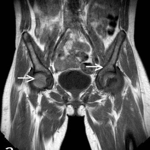
The patient initially underwent plain chest radiography, which was normal. Due to her pain and the tenderness, mostly at the left iliac bone, she was further evaluated with a pelvic MRI. The major question was whether the patient's symptoms were due to osteomyelitis or an acute infarction. On coronal T1W images (Fig. 1 a, b), the bone marrow had the characteristic low MR signal of sickle cell anaemia changes. On STIR images demonstrated massive bone oedema, more prominent at the left iliac bone and femora (Fig. 2 a, b). A small fluid collection was seen at the left illiopsoas muscle (Fig. 2 c). Following bone and soft tissue biopsies were negative. After intravenous therapy the patient was symptom-free and discharged home. Decrease of bone oedema was present on the STIR sequences (Fig 3 a, b) at the follow-up MRI approximately two months after the initial evaluation.
Discussion
Sickle cell disease (SCD) is a congenital haemoglobinopathy in which erythrocytes are dense, having a particular non-deformable shape. It may have different manifestations in numerous organs such as spleen, bones, eyes and central nervous system [1]. Vaso-occlusion crisis (VOC) is one of the most frequent complications in a SCD patient. The respective complication occurs due to microcirculation obstruction, which leads to local ischaemia and acute infarctions. Acute bone crises in SCD are precipitated by unknown mechanisms, although trauma, systemic infection, fever, stress, and other causes result in a really severe appearance of intense pain, tenderness, fever and local redness, same as in acute osteomyelitis (AO). These two pathologic entities have to be treated in a different way, although in most of the cases patients take large amounts of antibiotics. Radiographs are not helpful in acute bone marrow infarction (ABI), as they are usually normal during the initial phase of a VOC [1]. Additional radioisotope bone scan using a combination of radioisotopes can reliably detect areas but the specificity is low [2]. Ultrasound is mainly used as an interventional tool for ultrasound-guided biopsy. MRI is the gold standard imaging modality used, because it can detect the pathology of the bone marrow, soft tissues and intra-articular space, helping in the differential diagnosis and follow-up after treatment. Herein there is a large overlap between imaging findings of an ABI and AO, and therefore their differentiation might be quite a dilemma. Features that favour the diagnosis of osteomyelitis include involvement of the soft tissues and abscess formation. Also after intravenous contrast medium osteomyelitis may show a more geographic and irregular marrow enhancement, while acute infarcts demonstrates often a thin, linear rim enhancement. Although, contrast medium imaging can differentiate acute osteomyelitis from bone infarction, new studies demonstrate a potential role of MRI without contrast with equivalent diagnostic value [3]. Most patients recover from vaso-occlusive crises with no functional complications. Patients suffer chronic infarctions since early childhood [4]. However, when the infarction involves the epiphyses, it may give rise to joint effusions that are clinically similar to septic arthritis. Long-standing complications of infarctions at the epiphysis include short stature and kyphosis due to vertebral collapse. Avascular necrosis leads to faster and more severe disability, because infarctions in SCD are larger compared to other causes. In addition, hyperplasia of the bone marrow may cause osteopenia and growth disturbance [1, 4, 5]. Vaso-occlusive crisis could be due to acute infarction or osteomyelitis.
Differential Diagnosis List
Final Diagnosis
Acute bone infarcts
Liscense
Figures
MRI- Coronal T1-WI


MRI- STIR


Two-month follow-up, MRI- Coronal T1-WI

Seven-month follow-up, MRI- STIR


Imaging Findings
Based on the provided MRI images of the pelvis and hip joints, there are signal abnormalities in the bilateral proximal femurs (mainly involving the femoral heads and necks). On T1-weighted sequences, localized low or iso-signal intensity is observed, whereas T2-weighted sequences show areas of high or mixed signal intensity, with some lesions having linear or thin ring-like enhancement at the edges. No obvious extensive soft tissue swelling or abscess formation is evident, and no large effusion is seen in the joint space. Some images show scattered or patchy abnormal signals in the femoral heads, suggesting changes in the bone marrow structure. However, soft tissue involvement appears relatively limited, and there is no indication of widespread soft tissue destruction or notable abscess signals.
Potential Diagnoses
- Bone Infarction (Acute Bone Marrow Infarction/Vaso-occlusive Crisis): The patient has sickle cell disease (SCD), which predisposes to vascular occlusion and leads to local ischemic infarction. On imaging, characteristic findings may include abnormal signals in the femoral head or shaft, especially band-like or patchy high T2 signals, often showing thin or linear ring enhancement. These findings are consistent with SCD-related acute bone infarction described in the literature.
- Acute Osteomyelitis: Patients with SCD are overall more susceptible to infections, raising the possibility of osteomyelitis. In osteomyelitis, one often sees soft tissue swelling, obvious abscess formation, or necrotic tissue, and post-contrast scans typically show irregular marrow cavity enhancement, possibly accompanied by periosteal reaction. However, current imaging does not reveal clear abscess formation or extensive soft tissue inflammation, so further evaluation with clinical infection markers is necessary to rule it out.
Final Diagnosis
Combining the patient’s history of sickle cell disease, clinical symptoms (fever and bone pain), and MRI findings, the most likely diagnosis is acute bone infarction (bone marrow infarction caused by a vaso-occlusive crisis). Although osteomyelitis remains a differential diagnosis, the present imaging characteristics more strongly suggest ischemic damage secondary to a vaso-occlusive crisis.
Treatment Plan and Rehabilitation Program
Treatment Strategy:
- Conservative and Medical Management: Depending on the patient’s pain level and laboratory inflammation markers (after differentiating from osteomyelitis), consider the use of analgesics, sedatives, and other supportive treatments. If infection is suspected or if there is a high risk of infection, empirical antibiotic therapy can be used and adjusted according to microbiological findings.
- Transfusion and Hydration: In patients with sickle cell disease, blood transfusions can reduce the proportion of abnormal red blood cells and improve oxygen delivery. Maintaining adequate hydration can help alleviate vascular occlusion.
- Other Medications: Agents such as hydroxyurea can reduce the frequency of sickle cell crises. As necessary, other treatments targeting comorbid conditions (e.g., infection, thrombosis) should be considered.
- Surgical Indications: If, in the future, collapse of the femoral head or significant destruction of the joint occurs, leading to severe functional impairment, possible interventions include debridement, joint reconstruction, or joint replacement. Currently, conservative and medical management remain the primary approaches during the acute phase.
Rehabilitation and Exercise Prescription:
- Early Rehabilitation (Acute Phase): Focus on reducing weight-bearing and managing pain while maintaining joint range of motion. Within pain-tolerable limits, conduct simple active or passive hip joint movement exercises (e.g., flexion, extension, internal and external rotation) for 5–10 minutes, 2–3 times per day.
- Mid Rehabilitation (Symptom Alleviation Phase): Upon medical approval, gradually increase non-weight-bearing or partial weight-bearing exercises, such as walking with support or performing hip function exercises in an upright position for 10–15 minutes, 2–3 times per day, with a moderate increase in intensity.
- Late Rehabilitation (Functional Recovery Phase): Depending on the patient’s bone density and recovery status, progressively introduce weight-bearing activities, combining stretching exercises and mild resistance training (e.g., resistance bands) for 15–20 minutes, 3–5 times per week. Closely monitor pain levels and changes in joint mobility.
- Individualization and Safety: If the patient has significant osteoporosis, anemia, or compromised cardiopulmonary function, further reduce exercise intensity and extend rest periods to ensure safety. Warm-up and cool-down sessions should be done before and after each exercise session, avoiding abrupt high-intensity movements or sudden twisting.
Disclaimer
This report is based solely on the provided medical history and imaging data and is intended to serve as a reference for clinical diagnosis and treatment. It cannot replace in-person medical consultations or the definitive opinion of a qualified physician. Final diagnostic and treatment decisions should be made according to the patient’s actual condition and under the guidance of an appropriate specialist.
Human Doctor Final Diagnosis
Acute bone infarcts