Atypical non-ossifying fibroma


Clinical History
37-year-old man presented at the emergency department with a painful knee following a twisting injury.
Imaging Findings
AP and lateral radiographs demonstrate a 35x90 mm lucent lesion at the metadiaphysis of the right femur. It is central, with ill-defined, non-sclerotic borders and cortical erosion. Medial periosteal reaction is noted.
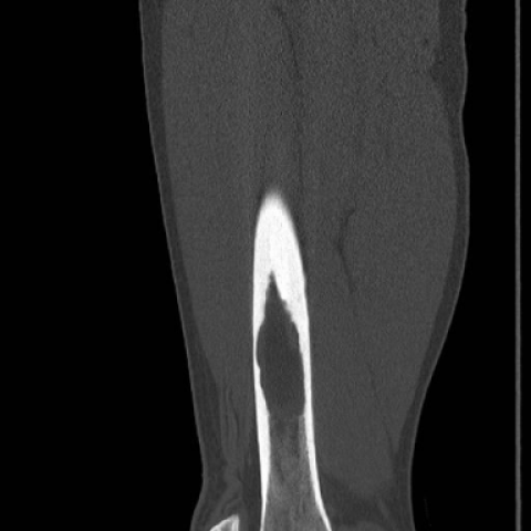
CT without contrast confirms an intramedullary lesion. No chondroid matrix. Periosteal reaction appears solid.
Discussion
Non-ossifying fibroma (NOF) is a benign [1] fibrous [2] lesion. It is histologically identical to fibrous cortical defect (FCD), but, by convention, is larger than 3 cm in its longest dimension [1]. It is rarely found in patients over 20 [3] and is twice as common in men [4]. NOF and FCD are extremely common: found in 33% of normal children and the most common benign skeletal lesions [1]. NOF is most often found near the knee: at the distal femur or proximal tibia [5]. It is thought to be a developmental defect [2] and originates at the metaphysis, growing into the diaphysis [3]. In 8% of cases, there are multiple lesions [6].
NOF is generally asymptomatic--an inconsequential finding found incidentally on imaging ordered for other reasons [4]. However, especially large lesions can cause chronic pain [1] and risk of pathologic fracture, particularly once they involve more than 50% of the bone's transverse diameter [4].
Radiographs are most often sufficient for diagnosis [6]. NOF classically appears as a well-circumscribed, eccentric radiolucency [7] in a metaphyseal or metadiaphyseal position around the knee [2]. When large, it can appear centrally [2]. It is usually ovular, with its long axis parallel to that of the bone, and it protrudes into the medullary cavity [6]. The appearance of its margins can vary, from indistinct to densely sclerotic, as can its number of loculi [4]. This is a result of its natural history: at first lytic, the lesion is eventually overcome by sclerosis, and then ossification from the diaphyseal side, followed by remodelling to normal bone [2, 3]. This sclerosis can be mistaken for fibrous dysplasia [8]. Periosteal reaction occurs only with a pathologic fracture involving the lesion [8]. Further imaging is not indicated unless surgery is required or the presentation is atypical, and, in these cases, CT is the preferred modality [6, 8]. CT allows for detailed assessment of size and volume [6] and can evaluate the integrity of the bony cortex [5].
NOF is slow-growing [8], and most will naturally heal over a period of 29-52 months from the end of adolescence [1]. No treatment is warranted [2]. In large defects, patients may be advised to avoid strenuous activity that may precipitate a fracture, which would then be treated with either reduction and immobilization or curettage and bone graft [4].
NOF is a common, benign lesion that can be mistaken for something more serious, particularly when it presents atypically.
Differential Diagnosis List
Final Diagnosis
Non-ossifying fibroma (confirmed by pathology)
Liscense
Figures
Radiographs


CT without contrast


Imaging Findings
Based on the provided knee X-ray and CT images, a relatively large, well-defined, eccentric lytic lesion is observed in the metaphyseal region of the distal right femur (near the knee joint). The lesion appears elliptical or quasi-elliptical in shape, with its long axis parallel to the diaphysis. A noticeably radiolucent area is visible within the lesion, and the local cortical bone appears slightly thinned yet remains mostly intact, without definite signs of cortical disruption. A certain degree of sclerotic margin is noted, with no obvious periosteal reaction and no clear evidence of soft tissue swelling or mass. Correlating with the axial and coronal CT images, the lesion is located within the medullary cavity and presents imaging characteristics consistent with a benign bone lesion.
Potential Diagnoses
- Non-ossifying Fibroma (NOF): Commonly found in the metaphysis of long bones, typically presenting as an eccentric, elliptical lytic lesion with or without a sclerotic rim. It can undergo gradual sclerosis over time. NOF is often seen in adolescents and individuals under 20 years old, but it may also occur in young adults or adults. Its imaging features are relatively consistent with this case.
- Fibrous Dysplasia: May exhibit “ground-glass” bone density changes. A sclerotic border can sometimes be present; however, on CT, the internal density often appears irregular and mixed, and the margins are typically less defined than those of a non-ossifying fibroma. While this case shows some sclerotic changes, on the whole, it is more suggestive of an NOF.
- Giant Cell Tumor of Bone: Commonly occurs in the epiphysis and can extend to the articular surface, typically affecting individuals between 20–40 years old. These tumors are often expansile and destructive, causing thinning or ballooning of the cortex, and may extend into the surrounding soft tissue. Although the lesion in this case is large, there is no significant cortical expansion, destruction, or extraosseous soft tissue mass, making this diagnosis less likely.
Final Diagnosis
Taking into consideration the patient’s age (37 years old), history (twisting injury followed by pain), and imaging findings (a typical eccentric, radiolucent lytic lesion with some degree of sclerotic margin, slightly thinned cortical bone without obvious destruction, and no periosteal reaction), the most consistent diagnosis is Non-ossifying Fibroma (NOF). Although NOF is more commonly seen in adolescents, it can also occur in adults. In this case, the lesion is relatively large, but there is no clear evidence of pathological fracture or malignant transformation.
Treatment Plan and Rehabilitation
Treatment Strategy:
Most non-ossifying fibroma lesions require no special intervention and can be managed with periodic follow-up to monitor for any lesion enlargement or worsening of symptoms. For patients with no significant bone destruction or risk of pathological fracture, conservative management is generally sufficient, including:
- Symptom Management: Short-term nonsteroidal anti-inflammatory drugs (NSAIDs) can be used to relieve pain or discomfort.
- Avoiding Excessive Weight-Bearing or High-Impact Activities: If the lesion is large and occupies more than 50% of the bone’s diameter, there is a potential risk of pathological fracture, and high-impact sports should be modified or reduced accordingly.
- Surgical Indications: Surgery (curettage, bone grafting, internal fixation) may be considered only if there is a significant risk of pathological fracture or if symptoms persist and cannot be managed conservatively.
Rehabilitation/Exercise Prescription Recommendations:
Rehabilitation exercises should follow a gradual and individualized approach. Begin with exercises that do not cause marked pain or discomfort, focusing on preserving joint range of motion and strengthening muscles while minimizing stress on the knee. The following FITT-VP principles can be used:
- Frequency (F): Engage in low-impact exercises (e.g., swimming, stationary cycling, core training) 3–4 times per week.
- Intensity (I): Start with low intensity and gradually increase resistance or duration; avoid strenuous running or jumping.
- Time (T): Each exercise session can last 20–30 minutes, with appropriate warm-up and cool-down.
- Type (T): Emphasize low- to moderate-intensity aerobic exercise combined with light resistance training (e.g., resistance bands or light weights) and avoid heavy-load squatting.
- Progression (P): Increase duration and intensity based on pain and fatigue levels. Once discomfort subsides, gradually add heavier loads under the guidance of a specialist or physical therapist.
Disclaimer
This report is solely for reference based on the current imaging and clinical information and cannot replace in-person clinical diagnosis and medical advice. If you have any further questions or experience any change in symptoms, please consult a specialist. In addition, laboratory tests or pathological examinations may be necessary to achieve a definitive diagnosis and treatment plan.
Human Doctor Final Diagnosis
Non-ossifying fibroma (confirmed by pathology)