

A 55-year-old man presented with worsening right heel pain but denied a history of trauma. Physical examination revealed a normal motion of the right ankle joint.
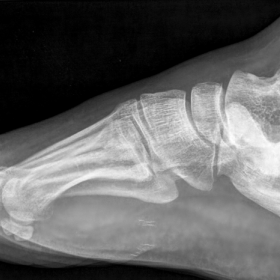
On plain film the calcaneus shows a sclerotic appearance with thickened trabecular pattern; no cortical breakthrough is present (Fig. 1). Bone scintigraphy shows intense radionuclide uptake only at the right calcaneus (Fig. 2).
MRI of the foot reveals a markedly unusual appearance of the calcaneus with trabecular disorganization and condensation; patchy marrow abnormality is evident. No evidence of malignancy (Fig. 3).
A follow-up performed each year for four years with annual MRI did not show any change of the findings (Fig. 4).
Paget's disease of bone is a metabolic disorder of unknown aetiology that is featured by irregular remodelling of bone leading to enlargement and deformity of osseous structures [1].
It is the second most common bone disorder after osteoporosis and affects an estimated 2-7% of persons aged 55 years or older [2] . In the majority of cases it is asymptomatic, and when present pain is the common symptom. The disease is infrequently monostotic, 85-90% of patients have the polyostotic variant [3].
The location of the feet is rare, 33% of the cases in the polyostotic variant and only 3% of the cases in the monostotic variant [4]. Bone scintigraphy, as in our case, is a valuable method in such situations, establishing the single localization of Paget's disease.
Radiographic features of Paget's disease correspond to the pathologic processes in the bone.
In early stages (osteolytic phase), active bone resorption appears as a radiolucent wedge or a stretched area with pointed margins that smashes both the cortical and cancellous bone as it advances along the bone. In the intermediate phase, bone destruction is characterized by bone formation, with the latter process tending to predominate. Bone remodelling appears as thickening of the cortex and coarse trabeculation of cancellous bone. In the cool or sclerotic phase, an increase of bone density occurs with enlargement and widening of the bone and cortical thickening, so that a demarcation between cortex and cancellous bone becomes difficult [5].
MRI is employed to show in a better way the cortical and intramedullary involvement, and to evaluate the extension of the disease into the soft tissues. On T1w sequences intermediate-to-low signal intensity is a common finding. On T2w sequences the signal may be either high, intermediate, or low, depending on the phase of the disease [5]. The most common pattern is dominant signal intensity in pagetic bone similar to the signal of fat; this pattern of involvement supposedly corresponds to long-standing disease and is seen in most patients. The second pattern likely corresponds to the early active phase, and in this stage the involved bone shows heterogeneous, relatively low T1 signal intensity and high T2 signal intensity. The least common pattern of signal intensity changes is seen in the cool or sclerotic phase when the affected bone shows low signal intensity on both T1- and T2w images, maybe related to the presence of compact bone or fibrous tissue [6].
Monostotic Paget’s disease of the calcaneus


Based on the provided right foot X-ray and MRI images, as well as bone scan results, the main radiological features are summarized as follows:
The overall radiological findings are consistent with a single focus of bone overgrowth, cortical thickening, coarsened trabeculae, and increased bone remodeling activity.
Taking into account the patient’s age (55-year-old male), chronic heel pain with no history of trauma, and radiological evidence of abnormal bone remodeling, the following diagnostic considerations are proposed:
Based on the patient’s age, clinical presentation (chronic heel pain), radiological findings (bone thickening, density changes, and rare monostotic involvement), and the bone scan results indicating a single site of elevated metabolic activity, the most likely diagnosis is monostotic Paget’s disease (calcaneus involvement). If there remains any clinical doubt, further tests such as serum alkaline phosphatase measurement or bone biopsy could be considered to confirm the diagnosis.
The following therapeutic strategies and exercise-based rehabilitation recommendations may be considered for this case:
These should be adapted to the patient’s overall condition and bone mass status, following the FITT-VP principle (Frequency, Intensity, Time, Type, Volume-Progression):
In cases of severe deformity causing functional impairment or refractory pain, surgical treatment (e.g., bone resection or corrective surgery) can be considered. For this patient, who currently has pain localized to the calcaneus, if conservative measures prove ineffective and bone deformity progresses, referral for a specialized surgical consultation may be warranted.
Disclaimer:
This report provides a reference analysis based on the available information and does not replace an in-person consultation or professional medical advice. Patients should seek personalized treatment and rehabilitation under the guidance of a qualified physician.
Monostotic Paget’s disease of the calcaneus