Subacute osteomyelitis (ECR 2013 Case of the Day)


Clinical History
A 51-year-old patient was referred to our University Medical Centre with the diagnosis of an osteosarcoma of the left upper arm. He had a 10-week history of pain and swelling of his left upper arm, which increased in the evening. He had no fever in this period.
Imaging Findings
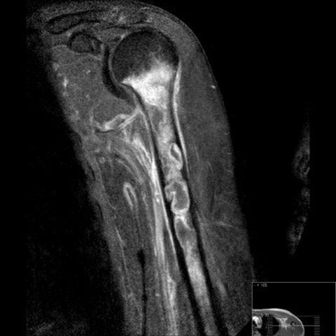
The conventional radiograph of the left upper arm showed a geographic to moth-eaten lobulated osteolytic lesion in the proximal meta-diaphysis of the humeral bone with a discrete periosteal reaction. No matrix was present (Fig. 1). Magnetic Resonance Imaging of the left upper arm was performed. These images showed an intramedullary lesion of at least 20 cm. The T2 weighted image with fat-suppression showed several well-demarcated collections with high signal intensity.
One of these collections showed the typical "penumbra sign" on the T1 weighted image without contrast, which consist of a lower signal intensity area in the centre and a high signal intensity rim (Fig. 2a). This high intensity rim was enhancing after gadolinium administration while the centre did not enhance, consistent with fluid/pus (Fig. 2b, c). Furthermore, multiple high signal intensity foci on the T1 weighted images without contrast were visualised, compatible with fat globules (Fig. 3a, b).
Discussion
Subacute osteomyelitis can be difficult to diagnose because the characteristic clinical signs and symptoms of acute infection can be absent. Patients are often not systemically ill, and laboratory results may be normal. Osteomyelitis can be secondary to sepsis, surgery or trauma. Its origin is typically located in the metaphysis. In direct trauma, as may be the cause in our patient, sinus thrombosis leads to stasis and infection.
The first step in imaging primary osteomyelitis is a conventional radiograph. Typically, it shows a mixture of soft tissue swelling, osseous destruction, multilamellar periosteal reaction and reactive sclerosis. Brodie’s abscess usually presents as a well-defined round or ovoid radiolucency [1]. These radiographic appearances of subacute osteomyelitis can be mistaken for malignant or benign bone tumours. Although a matrix is not formed in the presented lesion, an aggressive osteosarcoma cannot be excluded.
MRI is the most sensitive (approaching 100%) radiological modality to detect osteomyelitis, however, the specificity is much lower (approximately 80%) [2]. Several reports focus on specific MRI findings that may increase specificity. First the ‘penumbra sign’ is described as a characteristic MR feature for subacute osteomyelitis. This is thought to be helpful in differentiating subacute osteomyelitis from a neoplasm [3, 4]. Davies et al described the presence of fat globules as a specific sign for osteomyelitis. They presume that the presence of intra- and extramedullary fat globules is the result of septic necrosis and destruction of the normal lipocytes. The pattern of fatty signal may be diffuse or focal [5]. Although this sign is described in the acute form of osteomyelitis, we observed it in our patient in combination with the penumbra sign. This is probably due to an acute exacerbation of a subacute/chronic osteomyelitis. The diagnosis was confirmed with biopsy, which showed a chronic osteomyelitis with an acute component. The patient was referred for appropriate treatment of the infection; cleansing and antibiotic therapy.
Conclusion: Diagnosing osteomyelitis can be challenging on both conventional radiographs and MRI. However, knowledge of specific MR signs can help in differentiating a lesion from a malignant origin.
Differential Diagnosis List
Final Diagnosis
Subacute osteomyelitis of the left humeral bone.
Liscense
Figures
Coronal T1 weighted image; fat globules


AP conventional radiograph of the left upper arm

Axial T1 weighted image


Medical Analysis Report
I. Imaging Findings
1. X-ray Findings: Local bone destruction and sclerosis are observed in the proximal to mid portion of the humeral shaft on the affected side. Layered (multilayered) periosteal reaction is visible, along with marked soft tissue swelling.
2. MRI Findings: On T1WI, the lesion shows relatively clear boundaries with moderate to low signal intensity. A “penumbra sign” may be noted within the lesion, and scattered fat signals or fat droplet–like foci (indicating destroyed adipose tissue) can be seen on T2WI or fat-suppressed sequences. Contrast enhancement reveals marginal enhancement; in some regions, marked enhancement is observed, and inflammatory edema signals are present in the surrounding soft tissue.
II. Potential Diagnoses
Based on the imaging findings and the patient’s clinical symptoms, the possible diagnoses include:
- Subacute/Chronic Osteomyelitis:
∙ Imaging may show local bone destruction, sclerosis, periosteal reaction, and characteristic “penumbra sign” along with fat necrosis signals;
∙ Clinically, the patient presents with pain and swelling but lacks typical systemic signs of infection;
∙ The course is relatively long and may be related to trauma, bacteremia, or infection due to local injuries. - Malignant Bone Tumor (e.g., Osteosarcoma):
∙ Can also present with bone destruction and periosteal reaction (such as Codman’s triangle or onion-skin periosteal reaction);
∙ MRI may demonstrate a tumorous mass, often accompanied by a soft tissue component;
∙ Some osteosarcomas lack a clear bony tumor matrix, so suspicion of malignancy should remain high.
III. Final Diagnosis
Considering the patient’s history of local pain and swelling for over 10 weeks, the absence of significant fever or systemic infection signs, and the presence of a “penumbra sign” and fat droplet–like signals on MRI, a pathological biopsy confirmed “chronic osteomyelitis with acute exacerbation.” Therefore, chronic (subacute/chronic) osteomyelitis is more likely than a primary malignant bone tumor.
IV. Treatment Plan and Rehabilitation Plan
1. Treatment Plan:
∙ Surgical Debridement: For significant infectious lesions and possible necrotic bone, prompt surgical debridement is recommended to remove sequestrum and purulent material;
∙ Antibiotic Therapy: Based on pathogen testing, select sensitive antibiotics. Prolonged intravenous or oral antibiotic therapy (e.g., 4–6 weeks or longer) may be needed to control the infection;
∙ Supportive Therapy: Includes basic nursing care, nutritional support, and symptomatic treatment (e.g., pain management).
2. Rehabilitation Plan and Exercise Prescription (FITT-VP Principle):
∙ Type of Exercise: Initially low-load, non-weight-bearing or partial weight-bearing activities such as seated and standing joint movements, stationary bicycle (if feasible), and light resistance band exercises;
∙ Frequency: 3–5 times per week, adjustable according to the patient’s tolerance and infection status;
∙ Intensity: Begin with very light to moderate intensity (e.g., Borg Rating of Perceived Exertion 9–11), avoiding significant swelling or exacerbation of pain;
∙ Time: 15–30 minutes per session, or split into 2–3 segments of about 10 minutes each, gradually increasing over time;
∙ Progression: As the infection improves and pain/inflammation subsides, gradually increase load and resistance, following a step-by-step approach with regular follow-up;
∙ Volume: During the acute phase, strictly limit the activity volume to reduce local irritation and promote healing; in the chronic phase, gradually increase the training volume.
Throughout the rehabilitation process, the affected limb should be closely monitored. If local pain, redness, or systemic discomfort intensifies, training should be paused, and medical advice should be sought promptly. It is crucial to protect the affected limb from external impact or excessive load to prevent reinjury or worsened infection.
Disclaimer: This report serves as a reference analysis and cannot replace in-person consultation or the expert opinion of a professional physician. If you have any questions or need further evaluation and treatment, please consult a clinical specialist.
Human Doctor Final Diagnosis
Subacute osteomyelitis of the left humeral bone.