Soft tissue chondroma (ECR 2015 Case of the Day)


Clinical History
A 79-year-old man presented with pain and swelling at the plantar side of his left foot for several months. There was no history of acute trauma. His past medical history was unremarkable.
Imaging Findings
Radiography and magnetic resonance imaging (MRI), including post-gadolinium images, were performed.
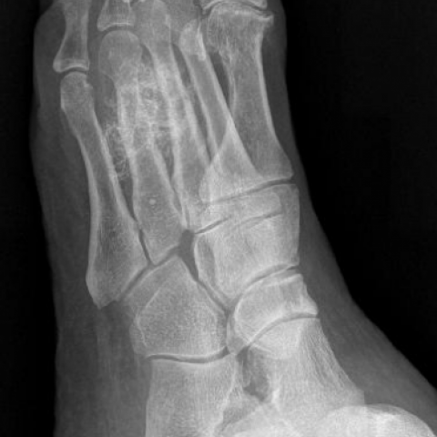
Radiographs show a distinct round lesion with ring and arc-like calcifications in the soft tissue at the plantar side of the left foot. There is also a consolidated fracture of the diaphysis of the fourth metatarsal.
MRI demonstrates a well-defined, lobulated lesion with a maximum diameter of 5 cm, without a clear connection to the metatarsals. The lesion is heterogeneous and has mainly a high signal intensity on both T1-weighted and T2-weighted images. After administration of gadolinium contrast material, peripheral rim enhancement is seen. There are intralesional areas of low signal intensity on all sequences, compatible with calcifications. No significant perilesional oedema is depicted on fat-suppressed proton-density weighted images.
Discussion
Background
Soft tissue chondromas are benign soft tissue tumours occurring in extraosseous and extra-synovial locations. [1] They constitute 1.5% of benign soft tissue tumours. [2] The most commonly accepted theory of tissue origin is synovial. [3] Soft tissue chondromas are predominantly composed of hyaline cartilage but can also contain osseous, fibrous and/or myxoid stroma. [1]
Clinical Perspective
The majority of patients are middle-aged, with an age range of 1-85 years. [2] There is a slight male predominance. [2] Most lesions arise in the hands and feet. [2-5] Patients usually present with a slowly growing soft tissue mass, occasionally with pain or tenderness. [2]
Imaging Perspective
Radiographically, soft tissue chondromas are well demarcated, lobulated lesions with central and peripheral calcifications, often curvilinear in nature. [2-5] Calcifications are radiographically visible in 33-70%. [2] Although they do, by definition, not originate from intraarticular synovium or periosteum, [1] remodelling, erosion, or sclerosis of nearby bone can occur. [2, 5] The MRI appearance is variable and probably related to the degree of calcified matrix. [3] The main role of MRI is to localize and determine the extent of the lesion. In our case, location and the presence of chondroid-like calcifications suggest soft tissue chondroma; synovial sarcoma, heterotopic ossification, and calcific myonecrosis are less probable. Synovial sarcoma may calcify in 30% of cases and is usually seen in patients of 30-40 years of age. The MRI enhancement pattern of synovial sarcoma is more pronounced than of soft tissue chondroma. In 50% of heterotopic calcifications due to myositis ossificans, there is a trauma history. Calcific myonecrosis shows typical plaque-like calcifications along the orientation of the muscle fibres. MRI showed no direct connection with bone, ruling out osteochondromatous lesions. [6] An extraskeletal chondrosarcoma, however, could not be ruled out by imaging alone. Therefore, biopsy was performed, showing cartilage and some bone, without signs of malignancy, confirming the diagnosis of a soft tissue chondroma. Notably, the presence of an old healed fracture of the fourth metatarsal may have been a coincidental finding. Yet, it has also been suggested that trauma may induce soft tissue chondroma formation. [7]
Outcome
Local excision was performed, which is usually curative. However, 15-25 % of soft tissue chondromas may locally recur. [2] Chondrosarcoma transformation has not been described. [1]
Take Home Message
A soft tissue chondroma is a rare benign soft tissue lesion, most frequently arising at the hands or feet. Radiography typically demonstrates a well-demarcated, lobulated soft tissue lesion with chondroid-like calcifications. MRI appearance of the lesion can be variable, but there is no osseous connection.
Differential Diagnosis List
Final Diagnosis
Soft tissue chondroma
Liscense
Figures
AP radiograph

Axial T1-weighted image

Axial fat-suppressed T1-weighted postgadolinium image

Axial T2-weighted image

Lateral radiograph

Sagittal T1-weighted image

Oblique radiograph
Radiological Findings
Based on the provided X-ray and MRI scans, the following can be observed:
- A well-defined lobulated soft tissue mass is seen within the plantar soft tissue of the left foot.
- On the X-ray, cartilage-like calcifications are observed within and around the mass, partially presenting as ring or segmented calcifications. No obvious bony connection or significant bone destruction is noted.
- MRI shows heterogeneous signal intensities on T1 and T2 sequences, with mixed high and low signals in certain areas. The mass margins are clear, with no definite involvement of bone.
- No significant invasion into adjacent structures is noted. An old healed fracture is visible in the fourth metatarsal.
Possible Diagnoses
Based on the patient's clinical presentation (several months of plantar pain and swelling without a clear history of acute trauma) and the imaging findings (cartilage-like calcifications within the soft tissue mass, no obvious bony connection or destruction), the following diagnoses are considered:
- Soft tissue chondroma
Characteristics: A benign cartilaginous tumor arising in the soft tissues, sometimes accompanied by calcification or ossification; commonly found in the hands and feet. Radiologically, it often appears as a lobulated soft tissue mass with cartilage-like calcifications. - Synovial sarcoma
Characteristics: Can exhibit calcification and typically occurs in individuals aged 30–40. On MRI, contrast enhancement is often more pronounced than in this case, and it can sometimes involve periarticular structures. - Heterotopic ossification / Myositis ossificans
Characteristics: Often follows trauma or surgery. In early stages, it appears as a soft tissue mass. Later, a more regular zone of ossification forms. The pattern of calcification differs from that of cartilage. - Calcific myonecrosis
Characteristics: Typically associated with severe trauma, multiple surgeries, or chronic ischemia. It manifests as band-like or layered calcification following the orientation of muscle fibers. - Extraskeletal chondrosarcoma
Characteristics: A more malignant entity that can show calcification. Histologically, typical features of chondrosarcoma may be present. It generally grows faster, accompanied by pain and more aggressive appearances on imaging.
Final Diagnosis
Considering the patient’s age, clinical symptoms (a slowly growing soft tissue mass with pain for several months), local imaging findings (a soft tissue tumor-like lesion with cartilage-like calcifications and no obvious bone destruction), and histology/pathology results (showing cartilaginous tissue and partial ossification with no clear signs of malignancy), the most likely diagnosis is:
Soft tissue chondroma.
This condition is generally benign, with malignant transformation being extremely rare. If pathological findings suggest malignancy, extraskeletal chondrosarcoma should be ruled out. In this case, there is currently no evidence of malignancy based on histological examination.
Treatment Plan and Rehabilitation
Based on the diagnosis and local symptoms in this case, the following recommendations are provided:
- Treatment Strategies
- Surgical excision: Complete excision is typically performed for soft tissue chondromas. Postoperative monitoring is necessary, as local recurrence rates can reach 15–25%, though distant metastasis or malignant transformation is extremely rare.
- If pain and functional impairment gradually subside after excision, regular follow-up is required, including imaging examinations to detect any local recurrence.
- For patients who are not suitable candidates for surgery or decline surgery temporarily, conservative treatment (e.g., temporary pain management and physical therapy) can be considered based on clinical symptoms. However, surgical treatment is recommended if the tumor significantly affects foot function or causes pronounced pain.
- Rehabilitation and Exercise Prescription
Postoperative rehabilitation should be adjusted according to the patient’s wound healing, pain level, and activity capacity. In general, it follows the gradual FITT-VP principle (Frequency, Intensity, Time, Type, Progression, and Individualization):
- Early Phase (1–2 weeks after surgery):
- Mainly focus on wound care and static muscle strength exercises, avoiding heavy weight-bearing on the foot.
- Within a safe range, perform gentle active movements of the ankle and toes to promote circulation.
- Frequency: 1–2 sessions per day, each lasting about 10–15 minutes.
- Mid Phase (2–6 weeks after surgery):
- Gradually increase localized weight-bearing activities, such as walking with braces or crutches.
- Emphasize foot joint range of motion and soft tissue stretching exercises to prevent adhesions or contracture of the plantar fascia and aponeurosis.
- Frequency: 3–4 sessions per week, each session 15–20 minutes, adjusted according to tolerance.
- Late Phase (6 weeks and beyond):
- Gradually transition to walking without aids and moderate weight-bearing exercises, continuing stable training as tolerated based on pain levels.
- Light balance and strength training under professional guidance can be introduced, such as resistance exercises for the small foot muscles or band exercises.
- One or two sessions of moderate-intensity exercises per week (e.g., swimming, cycling), each lasting 20–30 minutes, can improve foot endurance and overall cardiorespiratory fitness.
Throughout the rehabilitation process, close monitoring of foot pain and swelling is essential. In case of significant abnormalities or high-risk factors (such as fragile foot bones or poor cardiopulmonary function), prompt follow-up and adjustment of the training program are advised.
- Early Phase (1–2 weeks after surgery):
Disclaimer
The above report is a reference analysis based on the current information and cannot replace an in-person consultation or professional medical advice. If you have any questions or if your condition changes, it is recommended you promptly visit a hospital for further evaluation and treatment.
Human Doctor Final Diagnosis
Soft tissue chondroma