Camurati-Engelmann disease


Clinical History
A 26-year-old male patient presented at the emergency department after trauma to the right wrist, left shoulder and the head after an assault. He also mentioned long-standing limb pain, especially around the knees and ankles. Neurological examination was normal and there were no laboratory abnormalities.
Imaging Findings
There are no signs of recent trauma.
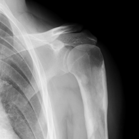
Plain radiographs show thickening of the diaphyses of the long bones (Figs. 1-4), including the distal femur, the tibia and the fibula (Figs. 3 and 4). These findings are bilateral and symmetric, with epiphyseal sparing (Figs. 3 and 4). Similar abnormalities are observed in the left proximal humerus (Fig. 1) and right distal radius and ulna (Fig. 2). There is minor cortical sclerosis in some metacarpal and metatarsal bones. The carpal bones, tarsal bones and phalanges of the hands and feet are normal (Figs. 2 and 4).
Brain CT shows no signs of acute trauma but reveals sclerosis and thickening of the cranial bones (Fig. 5). There is also narrowing of the optic canals (Fig. 5a).
Clinical presentation and radiographic abnormalities may correspond to Camurati-Engelmann disease.
The patient had a confirmed diagnosis of Camurati-Engelmann disease and a sister with the same condition.
Discussion
Camurati-Engelmann disease (CED), also known as progressive diaphyseal dysplasia, is a rare form of sclerosing bone dysplasia, [1, 2] belonging to the group of disturbance in intramembranous bone formation. [3] CED is an autosomal dominant disease with variable penetrance, [1, 2] in most cases related to mutations in the TGFB1 gene. [3, 4] About one person per million is affected. [5]
Phenotypical expression is highly variable and the age of onset is unpredictable. [4, 6] Although usually detected during childhood, [7] CED may have an indolent or quiescent course, [6] with cases of onset up to the sixth decade. [3] Common symptoms are limb pain, waddling gait, easy fatiguability and muscular weakness. [3, 4] If the skull base is involved, there may be symptoms of cranial nerve compression. [3, 5] Systemic manifestations, such as hepatosplenomegaly and bone marrow dysfunction are uncommon. [3]
Laboratory abnormalities are also uncommon. [3]
Radiological signs have a penetrance of 94%, [4] and precede clinical symptoms. [5] Bilateral and symmetric cortical thickening of the diaphyses of long bones is very characteristic. [1, 2] Both the periosteal and the endosteal side are affected, with concomitant broadening of the diaphyses and narrowing of the medullary cavity, suggesting that both osteoclastic and osteoblastic activities are disturbed. [4, 5] There may even be partial obliteration of the medullary cavity. [3] Metaphyses and epiphyses are generally spared because they are formed by endochondral ossification. [7]
CED is a progressive disease, becoming more prominent with age. [5]
It affects the long bones of the lower limbs more frequently than the upper limbs. [2, 3, 5] In some patients there is sclerosis of the skull, mandible, thoracic cage, vertebrae, pelvis, metacarpals and metatarsals. [3, 4, 5] Carpal and tarsal bones, as well as phalanges are usually spared. [3]
Increased osteoblastic activity is detected on scintigraphy before clinical or radiographic evidence of the disease. [3, 4]
Typical clinical presentation of CED is very useful in the differential diagnosis with other sclerosing bone dyspasia. [1] However, its rarity and variable course may complicate the diagnosis, requiring molecular analysis in some cases. [4]
Preferred treatment consists of corticosteroids, using the advantage of their effect of decreasing bone density. [2, 4] They improve both clinical and radiological abnormalities but have long-term side effects. [4, 8] NSAIDs only provide analgesia. [4]
Surgery may be needed for neurological decompression or to reduce medullary stenosis. [4, 8]
Differential Diagnosis List
Final Diagnosis
Camurati-Engelmann disease
Liscense
This work is licensed under a Creative Commons Attribution-NonCommercial-ShareAlike 4.0 International License.
Figures
Radiograph of the left shoulder
Radiographic study of the right hand


Radiographic study of the knees


Radiographic study of the feet


Brain CT


Medical Analysis Report
I. Imaging Findings
Based on the provided X-ray and CT images, the following main features are observed:
- Long Bone Diaphysis and Metaphysis: In both lower and upper limbs (e.g., femur, tibia, humerus), there is pronounced cortical thickening with bilateral symmetrical changes. The shaft diameter is widened, and the medullary cavity is narrowed to varying degrees. In some areas, it appears almost “filled in.”
- Shoulder, Wrist Joints, and Foot Bones: The cortical bone in the shoulder and wrist also shows some thickening, though less pronounced compared to the lower limbs. This aligns with the clinical observation that lower limbs are more noticeably affected than upper limbs. The tarsal and metatarsal bones of the foot are mostly normal or exhibit mild thickening; overall, there is cortical thickening but not a significant, widespread sclerotic appearance.
- Cranial CT: There is localized thickening of the skull base, but no obvious signs of narrowed neural foramina or clear evidence of cranial nerve compression at this point.
- Soft Tissue and Fracture Status: No marked soft tissue swelling or acute fracture signs are seen. There may be a possibility of mild soft tissue contusion from trauma, but no evident fracture lines or significant dislocation.
II. Differential Diagnosis
Considering the patient is a 26-year-old male with long-term limb pain (especially around the knee and ankle joints), radiological findings of “bilateral symmetrical cortical thickening, narrowed medullary cavity,” and no significant laboratory abnormalities, the following differential diagnoses are proposed:
- Camurati-Engelmann Disease (Progressive Diaphyseal Dysplasia): Classic manifestations include symmetrical cortical thickening of the long bones, more pronounced in the lower limbs, with common clinical symptoms such as limb pain, fatigue, and abnormal gait. Most patients show no significant laboratory abnormalities. Diagnosis mainly relies on clinical presentation, radiological findings, and genetic confirmation.
- Osteosclerosis (e.g., Sclerosing Bone Dysplasia, Osteopetrosis): Various forms of bone sclerosis (e.g., resistant osteodysplasia, osteoma-like lesions) can also lead to cortical thickening. However, these often present as focal lesions or widespread systemic sclerosis, frequently involving the skull and pelvis, with somewhat different clinical profiles.
- Hyperostotic Conditions (e.g., Bone Overgrowth, Bone Island): These typically lack the widespread, bilateral symmetrical cortical thickening of long bones or may only show limited localized thickening, making it difficult to explain the patient’s chronic, multi-site pain symptoms.
III. Final Diagnosis
Combining the patient’s age, long-term lower limb pain, the classic imaging presentation of symmetrical cortical thickening and narrowing of the medullary cavity, along with generally normal clinical and laboratory findings, the most fitting diagnosis is:
Camurati-Engelmann Disease (CED, Progressive Diaphyseal Dysplasia).
For more precise confirmation, genetic testing (TGFB1 gene mutation) may be performed to support the diagnosis.
IV. Treatment Plan and Rehabilitation
-
Medication:
- Glucocorticoids: These can help reduce bone density, thereby alleviating clinical symptoms and radiological changes, but long-term side effects of steroids (e.g., osteoporosis, increased infection risk) must be monitored.
- Non-Steroidal Anti-Inflammatory Drugs (NSAIDs): Primarily used for pain relief in this condition, with limited impact on the progression of cortical thickening.
-
Surgical Intervention:
- If cranial or spinal canal stenosis causes nerve compression symptoms (e.g., cranial nerve dysfunction or spinal cord compression), decompression surgery may be considered.
- In cases of significant medullary cavity narrowing in the long bones, severe pain, or hematopoietic impairment, surgical interventions (such as medullary canal widening) can be evaluated.
-
Rehabilitation and Exercise Prescription:
In the absence of acute fractures or severe neurological symptoms, patients should be encouraged to engage in appropriate exercise programs, while taking into account the potential pain or muscle weakness caused by bone thickening. A gradual FITT-VP approach is recommended:
- Type of Exercise (Type): Low-impact aerobic exercises (e.g., swimming, stationary cycling, elliptical trainer) and moderate resistance training focusing on lower limb flexion-extension and core stability exercises.
- Frequency (Frequency): 3-4 times per week; initially, 2-3 times per week may be sufficient depending on tolerance.
- Intensity (Intensity): Start with low to moderate intensity. Monitor heart rate or perceived exertion (RPE), and avoid excessive weight-bearing or high-impact exercises. If notable pain or discomfort occurs, adjust accordingly.
- Time (Time): Initially 20-30 minutes per session, gradually increasing to 40-60 minutes. Consider dividing sessions to prevent fatigue.
- Progression (Progression): Gradually increase intensity or duration based on subjective pain levels and muscle strength assessments. If bone or joint discomfort arises, reduce the training load and consult a physician or rehabilitation therapist.
- Volume and Pattern (Volume & Pattern): Conduct periodic evaluations of exercise capacity and pain levels (every 4-6 weeks) and adjust the exercise plan under professional guidance.
If trauma to the wrist, shoulder, or head has resulted in soft tissue or joint injuries, recovery should be assessed to ensure a safe exercise regimen. Appropriate protective gear or physical therapy may be used as needed.
Disclaimer: This report is intended for reference only and cannot replace in-person diagnosis or professional medical advice. If you have any questions or if symptoms worsen, please seek medical attention promptly.
Human Doctor Final Diagnosis
Camurati-Engelmann disease