Distal Humerus Physeal Separation


Clinical History
A three-year-old child presented to the emergency department following an unwitnessed fall down a slide at an indoor playpark, sustaining a closed injury to the left elbow. Examination revealed a deformed, tender elbow in extension, with diffuse swelling. Further assessment of the circumstances ruled out non-accidental injury.
Imaging Findings
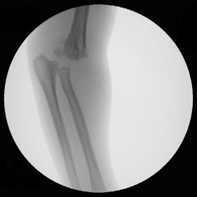
Radial axis in line with the ossified capitellum. The epiphysis, radius and ulna are displaced posteromedially.
Discussion
Distal humerus physeal separation (DHPS) is an injury that occurs in young children due to blunt trauma to the elbow joint. This injury is frequently associated with non-accidental injury (up to 50% [1]). It is therefore essential that the clinician assessing the injured child keep this diagnosis in mind. The injury may also be sustained following a fall from a height or from rotating forces through the elbow joint.
This injury is frequently mistaken for elbow dislocation which accounts for only 3-6% [2] of elbow injuries in all children. DHPS presents radiographically with subtle features, however close inspection reveals that the radial axis is inline with the capitellum ruling out a dislocation of the olecranon-trochlear articulation. The epiphysis, radius and ulna are displaced posteromedially which may be the only feature in the very young child with a non-ossified capitellum.
Anteroposterior, lateral and oblique view gives an assessment of the nature of the injury. It may prove challenging to obtain optimal views on plain imaging, especially in the young child. If the diagnosis is in doubt static or dynamic imaging may be required, under anaesthesia if necessary.
The treatment of this injury is closed reduction and internal fixation, ideally within four hours of presentation to the emergency department. This fracture healed clinically and radiologically with a satisfactory return of elbow function.
Detection of this injury is important due its significant association with non-accidental injury. Early recognition may lead to improved outcomes for the child, as growth abnormalities may be a complication of this physeal injury.
Differential Diagnosis List
Final Diagnosis
Distal Humerus Physeal Separation
Liscense
This work is licensed under a Creative Commons Attribution-NonCommercial-ShareAlike 4.0 International License.
Figures
Left elbow intraoperative (oblique view)

Left elbow AP view

Left elbow intraoperative (lateral view)

Medical Imaging Analysis Report
1. Imaging Findings
Based on the provided left elbow X-ray, there are visible indications of separation between the distal humeral physis and the diaphysis. Notable swelling is observed around the elbow joint, with the most prominent changes centered near the distal humerus physis. On the lateral view, the radius and ulna appear slightly displaced posteriorly and medially. It is noteworthy that, at certain angles, the radius still aligns with the capitulum (which is not fully ossified), suggesting that this is not merely a simple elbow dislocation. The soft tissue swelling and surrounding tissue shadows are moderately diffuse. Additionally, because the patient is very young and the ossification centers are not fully developed, close attention must be paid to subtle signs of physeal separation or fracture lines during imaging evaluation.
2. Potential Diagnoses
- Distal Humerus Physeal Separation (DHPS): In cases of elbow trauma in young children, physeal plate separation should be considered first, especially when external force leads to separation between the distal humeral epiphysis and the diaphysis.
- Posterior Elbow Dislocation: Although more common in older children, posterior displacement may appear in younger children with unclear imaging findings. However, in this case, good alignment of the radial head with the capitulum at certain angles reduces the likelihood of a simple posterior dislocation.
- Humeral Condylar Fracture: This includes fractures involving the lateral or medial condyle. On X-ray, one typically observes a clear fracture line or fragment displacement, which differs from a simple physeal separation.
The above differential diagnoses are primarily based on common pediatric elbow injuries and their distinct X-ray characteristics. Given the patient's age, limited protective reflexes, and relatively immature ossification centers at the elbow, physeal separation is often a primary consideration.
3. Final Diagnosis
Considering the patient’s age (three years old), the mechanism of injury (fall from a slide), and the imaging findings (physeal separation at the distal humerus, with overall posterior and medial displacement of the radius and ulna), the most likely diagnosis is: Distal Humerus Physeal Separation (DHPS).
4. Treatment Plan and Rehabilitation
4.1 Treatment Plan
- Closed Reduction and Internal Fixation: Such physeal separations often require closed reduction under general anesthesia or sedation, followed by temporary internal fixation with Kirschner wires or a small plate to align the physis and ensure stable healing. Early intervention (ideally within 4 hours after injury) is recommended for the best outcome.
- Postoperative Casting or Splinting: After confirming proper reduction, a cast or splint is usually applied to limit movement and promote bone healing.
4.2 Rehabilitation Plan
In preschool-aged children, the primary rehabilitation goal is to restore range of motion and muscle strength gradually. If necessary, simple functional training can be introduced after fracture healing. Because pediatric fractures tend to heal relatively quickly, it is important to balance protecting the growth plate with restoring joint function.
- Early Fixation Period (Weeks 1-2): Immobilization is the main approach during this phase. Avoid unnecessary limb movements. Monitor distal blood circulation and sensory function.
- Mid-/Late Fixation Period (Weeks 3-4): Once a physician has assessed that the fracture is stable, simple passive movement (e.g., gentle flexion and extension of the elbow) can be introduced safely.
- After Removal of Fixation (Week 4 and Beyond):
- Frequency: Perform joint mobility exercises 2-3 times a day.
- Intensity: Increase activity gradually, stopping before significant pain occurs.
- Time: Each session lasts about 5-10 minutes, gradually extending as recovery progresses.
- Type: Begin with passive or assisted exercises, progressing to active movements; focus on elbow flexion/extension and forearm rotation.
- Progression: As elbow function improves, gradually increase the range of motion and the number of repetitions. Light resistance exercises, such as lifting small toys, may be introduced in the later stages of recovery.
During the entire rehabilitation process, pay close attention to the child's subjective pain response, schedule regular follow-up imaging, and carry out exercises under the guidance of a professional rehabilitation therapist or orthopedic physician.
5. Disclaimer
This report provides a reference analysis based on available information and does not replace an in-person consultation or professional medical opinions. If there are any concerns or worsening symptoms, please seek medical attention promptly for further evaluation and treatment.
Human Doctor Final Diagnosis
Distal Humerus Physeal Separation