Preoperative digital planning in adult developmental dysplasia of the hip


Clinical History
A 64-year-old woman was investigated for surgical treatment of developmental hip dysplasia. Her complaint was bilateral hip pain and restriction in daily activities for the past two years. She had two deliveries and managed to compensate her disabilities before. The patient has short stature, hyperlordosis and equal lower limb length.
Imaging Findings
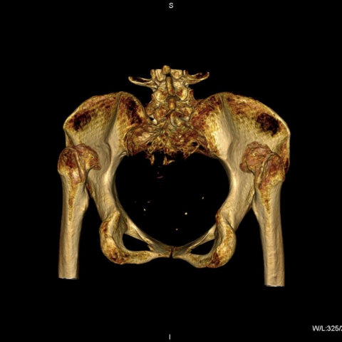
Digital pelvic radiography (Fig. 1) revealed the Crowe type IV bilateral developmental dysplasia of the hip (DDH). [1] Radiographic imaging of the pelvis enabled virtual preoperative templating (Fig. 2), although preoperative planning needed more sophisticated imaging techniques. Multiplanar CT of the pelvis was performed to further investigate bone stock and perform morphometric measurements of bony landmarks of the pelvis and femora (Fig. 3) MDCT scan of the pelvis provided accurate position of dislocated femoral heads relative to real acetabuli (Fig. 4). Essentially, 3D-CT scan showed bilateral high unsupported posterior hip dislocation, with exact relation between deformed femoral heads, iliac wings and true acetabuli. On both iliac wings two neoacetabuli were found, indicating an evolution of hip dysplasia (Fig. 5).
Discussion
Developmental dysplasia of the hip (DDH) may range from hardly detectable acetabular dysplasia to immensely malformed and highly dislocated hip.
The incidence of established developmental hip dislocation ranges between 1.5 and 20 per 1000 newborns. [2] Children whose parents had DDH are 10 times more prone to express DDH compared to their peers whose parents did not. [3]
Adolescent and adult DDH exists in two forms, those that were previously treated, and some that were left untreated, leading to premature hip osteoarthritis and groin pain. Almost 80% of total hip replacements (THR) performed in women and 15% in men are related to some degree to hip dysplasia. [4] Due to inefficient abductor musculature these patients limp or have a waddling (Trendelenburg) gait. [5]
Radiographic findings of DDH include shallow, mostly retroverted acetabulum and insufficient coverage of the femoral head, an increased femoral anteversion with shortened femoral neck and coxa valga. [6] Crowe classification is widely accepted in addressing THR for DDH, although frequently it is not a helpful tool for pre-operative planning. [1, 7] It is based on the extent of proximal migration of the femoral head addressing the ratio between the vertical distance of interteardrop line and the femoral head-neck junction (b) and the pelvic height (a); (type I < 50% subluxation or proximal dislocation <0.10; type II >50% and 74% subluxation or proximal dislocation 0.10 to 0.15, type III > 75% and 99% subluxation or proximal dislocation 0.16 to 0.20, type IV > 100% or complete dislocation >0.20). [1, 7] Eftekhar and Hartofilakidis proposed a radiologic classification depending on the grade of femoral head dislocation. The severity of DDH was divided into three and four stages, ranging from dysplasia to complete dislocation. [8, 9] CT studies confirmed a wide variety of deformities within the same Crowe grade. [10] MDCT provides valuable information regarding the type of deficiency and degree of acetabular dysplasia and may be used for 3D computerized preoperative planning. [11, 12] Preoperative digital planning has proven useful in joint replacement surgery allowing surgeons to choose an appropriate implant from the database [13, 14]
The patient underwent one stage modular total hip arthroplasty of the right hip with subtrochanteric shortening and realignment osteotomy according to preoperative digital templating (Fig.6a, b).
Take home message:
Digital preoperative planning in addressing total hip replacement in DDH improves the procedure's precision and its outcome, ensures the required implants are available, minimizing the costs and complications.
Differential Diagnosis List
Final Diagnosis
Developmental dysplasia of the hip (luxatio coxae congenita bilateralis)
Liscense
This work is licensed under a Creative Commons Attribution-NonCommercial-ShareAlike 4.0 International License.
Figures
Plain X-ray of the pelvis

Preoperative radiographic images of the pelvic region with virtual planning

MDCT scan of the pelvis – posterolateral view

Axial CT images of the pelvis

Postoperative images of the pelvic region

MDCT scan of the pelvis - PA view
Medical Imaging Analysis Report
I. Imaging Findings
Based on the provided pelvis anteroposterior (AP) X-ray and CT three-dimensional reconstruction images, the following main features are observed:
- Acetabular Dysplasia: Both sides show shallow acetabula with overall inadequate coverage, resulting in a large portion of the femoral head being exposed. Partial sclerosis of the bone and uneven joint space can be seen.
- Changes of the Joint Surface: Due to long-term abnormal stress, local osteophyte formation can be seen, and the joint space is narrowed, suggesting degenerative changes.
- Femoral Changes: Significant morphological changes of the proximal femur are noted. The femoral neck is relatively short and shows a certain degree of external (anterior) inclination. A mild deformity of the femoral head is seen.
- Crowe Classification: Based on measurement results (B/A = 0.38) and reference standards, the hip dislocation extent exceeds 0.20, indicating Crowe Type IV.
- Other Findings: The overall structure of the pelvis is relatively small, with no obvious abnormal soft tissue proliferation. CT axial measurements show varying degrees of abnormal anteversion angles and acetabular coverage on both sides.
II. Potential Diagnoses
Combining the imaging findings and the patient’s clinical history—a 64-year-old female with long-term hip discomfort and restricted mobility—the following diagnoses or differential diagnoses are considered:
- Developmental Dysplasia of the Hip (DDH) with Secondary Osteoarthritis: Based on the patient’s past history (abnormal weight-bearing in childhood, limping in adulthood, hip pain) and the marked dysplasia on imaging, DDH is likely. Obvious degenerative changes have already occurred, making this the most probable diagnosis.
- Secondary Degenerative Changes with Possible Femoral Head Necrosis: Femoral head necrosis typically shows collapse of the femoral head and disorganized trabeculae, but the current imaging findings primarily indicate congenital dysplasia. Signs of necrosis are not prominent, thus it is of secondary consideration.
- Inflammatory Joint Disease (e.g., Rheumatoid Arthritis): Usually presents with bilateral symmetrical synovitis and bone erosion, but the current imaging findings lack typical systemic or synovial changes, making this less likely.
III. Final Diagnosis
Based on the patient’s age, clinical symptoms (bilateral hip pain, limited range of motion), past medical history (childhood developmental issues not corrected promptly), and the above imaging findings (poor acetabular coverage, Crowe Type IV, etc.), the most likely diagnosis is:
Developmental Dysplasia of the Hip (Crowe Type IV) with Secondary Osteoarthritis.
The patient has undergone a “one-stage adjustable total hip arthroplasty with proximal femoral osteotomy” on the right side, which aligns with the treatment strategy for this diagnosis.
IV. Treatment Plan and Rehabilitation
1. Overview of Treatment Strategy
- Surgical Treatment: For patients with chronic pain and significantly impaired joint function—especially Crowe Type IV DDH—total hip replacement is an effective option. In cases of significant proximal femoral deformity, proximal femoral or subtrochanteric osteotomy may be considered to correct limb alignment and reduce soft tissue tension.
- Conservative Treatment: If activity is still tolerable or if the patient does not meet surgical criteria, short-term use of pain medication, joint protection devices, and physical therapy may be considered. However, such measures often have limited effectiveness in severe cases.
2. Rehabilitation/Exercise Prescription
Postoperative rehabilitation and exercise should be gradual and individualized, focusing on improving hip stability and restoring muscle strength and joint range of motion. The following plan may be considered:
- Frequency: 3–5 times per week.
- Intensity: Early postoperative rehabilitation should involve low-intensity exercises (e.g., non-weight-bearing exercises, partial weight-bearing under assisted devices), progressively transitioning to moderate-intensity activities (e.g., cycling, swimming).
- Time: Each session should be based on tolerance, starting with 10–15 minutes in the early stage, gradually increasing to about 30 minutes.
- Type:
- Joint Range of Motion Exercises: Initially, combine passive and active-assisted range of motion exercises, avoiding excessive adduction or flexion.
- Muscle Strength Training: Focus on strengthening the muscles around the hip (including the iliopsoas, gluteus medius, and gluteus maximus), such as straight leg raises and hip abduction with resistance bands.
- Aerobic Exercise: Options like swimming or using a stationary bike can enhance cardiovascular endurance while minimizing joint stress.
- Volume & Progression: Increase weight-bearing and exercise duration gradually, according to postoperative healing and functional assessments. Evaluate every 1–2 weeks and adjust the training plan accordingly.
Special Precautions: As the patient is older and may have osteoporosis or other comorbidities, closely monitor postoperative pain, swelling, and joint stability, and consult a physician or rehabilitation therapist promptly. Avoid high-impact, sudden power, or extreme twisting movements.
V. Disclaimer
This report is a reference analysis based on the provided medical history and imaging data and does not replace an in-person consultation or professional medical advice. Final diagnosis and treatment plans should be determined by a specialist physician, considering the patient’s clinical presentation and subsequent test results.
Human Doctor Final Diagnosis
Developmental dysplasia of the hip (luxatio coxae congenita bilateralis)