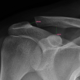


Pain at the right shoulder region following recent trauma.
Plain radiography: widened acromio-clavicular distance, measuring > 8mm while coraco-clavicular distance < 13mm is maintained.
MR: Sagittal T2 and coronal STIR images: thickening and oedema signal of the superior acromio-clavicular ligament.
Acromio-clavicular joint (ACJ) is a synovial joint. The distal clavicle articulates with the acromion process, with a small fibrocartilage articular disc in between and hyaline cartilage lining their articulating surfaces. The main joint stabilizers are: the deltoid and trapezius muscle (dynamic stabilizers) and the coraco-clavicular, acromio-clavicular and coraco-acromial ligaments (static stabilizers). [1, 2]
The coraco-clavicular ligament is an important stabilizer of the ACJ. It consists of two parts: the conoid ligament (posteromedial and triangular) and the trapezoid ligament (antrolateral and quadrilateral). The acromio-clavicular ligament is composed of superior, inferior, anterior and posterior components, the superior acromio-clavicular ligament is the strongest part and considered as thickening of the joint capsule. It merges with deltoid and trapezius muscle fibres. [1, 2]
On normal plain radiography, AP view, the acromio clavicular distance measures 1-3 mm and the coraco-clavicular distance measures 11-13 mm. Widening of the former exceeding 7 mm and/or the latter exceeding 13 mm indicates ACJ instability. Weight bearing (stress) and bilateral comparative views are helpful in some cases. The role of MRI is to illustrate ligament injury when radiography is normal or inconclusive. It allows more accurate staging of the ACJ injury according to Rockwood classification. [1, 2]
Rockwood classification is a grading system for ACJ injury which considers multiple factors as detailed later in text. The ACJ injuries are graded from 1 to 6, which influences the choice of management plan. Grades 1 & 2 are managed conservatively, while grades 4 to 6 are managed operatively. Grade 3 injury management is tailored for every patient. [3]
Grade 1: Partial tear of acromio-clavicular ligament.
Grade 2: Complete tear of acromio-clavicular ligament. Partial tear of coraco-clavicular ligament. Disrupted joint capsule. Minimal detachment of deltoid and trapezius fibres. Superior clavicular displacement < 50 %.
Grade 3: Complete tear of acromio-clavicular ligament & coraco-clavicular ligament. Disrupted joint capsule. Significant detachment of deltoid and trapezius fibres. Superior clavicular displacement 100 %.
Grade 4: Complete tear of acromio-clavicular ligament & coraco-clavicular ligament. Disrupted joint capsule. Significant detachment of deltoid and trapezius fibres. Posterior clavicular displacement.
Grade 5: Complete tear of acromio-clavicular ligament & coraco-clavicular ligament. Disrupted joint capsule. Significant detachment of deltoid and trapezius fibres. Superior clavicular displacement > 100 %.
Grade 6: Complete tear of acromio-clavicular ligament & coraco-clavicular ligament. Disrupted joint capsule. Significant detachment of deltoid and trapezius fibres. Inferior clavicular displacement.
Acromio-clavicular joint injury (grade I)
This work is licensed under a Creative Commons Attribution-NonCommercial-ShareAlike 4.0 International License.


Based on the right shoulder X-ray and MRI images provided by the patient, the main findings are as follows:
Based on the above imaging findings and the history of trauma (right shoulder pain after injury), the potential diagnoses include:
Considering the patient’s trauma history, clinical symptoms, and imaging characteristics (significant AC joint widening, coracoclavicular distance at the high normal value, and MRI suggesting partial or complete ligament tears), the most likely diagnosis is: AC joint injury, Rockwood Classification Grade II (complete or nearly complete tear of the AC ligament, partial tear of the CC ligament, with clavicle displacement not exceeding 50%).
Since the coracoclavicular distance remains within the upper normal limit (12.8 mm) without a clear increase beyond normal, and there is no marked superior displacement of the clavicle, Grade II injury features appear to be more fitting.
Rehabilitation should follow a gradual, individualized approach (the FITT-VP principle) to protect the injured area and progressively restore joint function and muscle strength:
If the patient has compromised bone quality or reduced cardiopulmonary function, they should increase training volume cautiously under the supervision of a professional rehabilitation therapist or physician, while closely monitoring for adverse effects. In case of significant pain or discomfort during exercise, the program should be adjusted promptly.
This report is a reference analysis based on the current imaging and medical history and does not replace in-person consultations or the opinions of professional physicians. If symptoms worsen or there is any uncertainty, please seek medical attention promptly.
Acromio-clavicular joint injury (grade I)