MR arthrography of SLAP X tear with rotator interval tear and biceps tendon rupture


Clinical History
A young athlete (Jiu-jitsu player) presented with pain and weakness in the right shoulder 3 months after trauma sustained during a competition. Physical examination revealed severe pain on abduction and inability to adequately flex the biceps muscle.
Imaging Findings
MR arthrography was performed for better delineation of lesions. Axial, sagittal and coronal images confirmed a large superior labral tear extending from 10 o’clock to 2 o’clock. The labrum was completely detached from the underlying glenoid bone with contrast noted between these 2 structures. The detached labrum was also split into fragments.
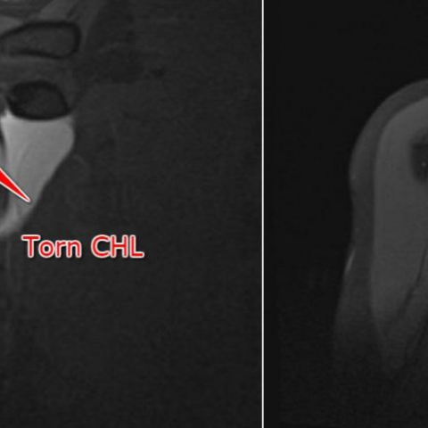
The rotator interval structures were also involved. Partial tear of the superior glenohumeral ligament was noted with fraying of the fibres. A large tear was also visualised in the coracohumeral ligament best seen on the coronal and sagittal images.
There was also rupture of the long head of biceps tendon with detachment from the biceps-labral complex. The tendon was also retracted into the bicipital groove.
The combination of findings were suggestive of a SLAP X tear and the findings were confirmed on arthroscopy.
Discussion
The superior labrum, rotator interval structures and the intra-articular portion of the long head of biceps tendon are some of the most difficult structures to evaluate by both MRI and arthroscopy. MR Arthrography has become the imaging modality of choice to evaluate these areas over the last decade [1, 2, 3]. In our institution, we use a mixture of 0.2 ml gadolinium, 10 ml iodinated contrast and 10 ml lidocaine to inject either via CT or ultrasound guidance.
Knowledge of anatomy and variants are critical to diagnosing lesions in these areas. The normal superior labral variants that simulate tears are noted from 11 - 3 o’clock. These include sublabral foramen, sublabral recess and Buford complex [1, 4]. The rotator interval consists of the capsule, superior glenohumeral ligament and the coracohumeral ligament between the subscapularis and supraspinatus tendons. It play an important role in preventing inferior and posterior translation at the gleno-humeral joint [5, 6]. These structures also form the biceps pulley while crossing over the biceps tendon and protect the tendon from dislocation [3]. High quality thin section arthrogram images are needed to demonstrate these structures effectively.
SLAP tears were originally classified into 4 types by Snyder depending on the extent of the labral tear and involvement of the adjacent biceps tendon. Over time, the classification has extended to 10 sub-types based on the extent of involvement of adjacent structures [1, 4]. Beltran first described the SLAP X tear in 2000 as a superior labral tear which not only involves the origin of the biceps tendon but also extends to involve the rotator interval structures (CHL and SGHL) with articular side abnormalities as seen in our case. This tear is important to recognise as it requires surgical management.
There are multiple other lesions that can involve the rotator interval and the biceps [7]. These include post-traumatic biceps pulley tears which lead to subluxation/dislocation of the biceps tendon. The tendon can also be affected by partial/complete tears and tendinitis. Adhesive capsulitis is manifested by thickening of the rotator interval capsule and ligaments with loss of normal fat and difficulty in intra-articular contrast injection.
Our case demonstrated a SLAP X tear with rupture of the biceps tendon. Due to the multiple subtypes of SLAP lesions, it is essential to describe the lesions in clock-wise distribution and the additional structures involved. Early operative repair followed by rehabilitation is essential [8].
Differential Diagnosis List
Final Diagnosis
SLAP X tear with rotator interval tear and biceps tendon rupture
Liscense
This work is licensed under a Creative Commons Attribution-NonCommercial-ShareAlike 4.0 International License.
Figures
MR Arthrogram images


Annotated images showing comparison with a normal study


Radiological Analysis Report
I. Radiological Findings
Based on the MR arthrography (MR Arthrography) sequences of the patient’s right shoulder, significant signal abnormalities are observed in the superior labrum region. The specific findings are as follows:
- The superior labrum (especially at the 11 to 1 o’clock position) shows disrupted structural continuity and increased signal intensity, indicating signs of tearing.
- The contrast agent injected into the glenohumeral joint between the glenoid and humeral head is seen infiltrating between the labrum and its attachment, increasing the suspicion of a tear.
- The long head of the biceps tendon appears incomplete as it enters the joint capsule; a localized discontinuity suggests a rupture of the tendon fibers or a high likelihood of such.
- At the humeral glenoid tubercle, partial joint fluid or contrast agent diffuses into the rotator interval (between the subscapularis tendon anteriorly and supraspinatus tendon superiorly), suggesting possible injury to the anterosuperior shoulder region and relevant ligamentous structures (such as the superior glenohumeral ligament and coracohumeral ligament).
- No definitive large-scale tear is seen in the surrounding tendons (supraspinatus, infraspinatus, and subscapularis); however, slight tenosynovial effusion signals can be observed.
II. Potential Diagnoses
Taking into account the patient’s age, history of jiu-jitsu practice, mechanism of injury, and clinical presentation (post-traumatic shoulder joint pain, aggravated by abduction, decreased biceps strength), the following possible diagnoses are considered:
-
SLAP (Superior Labral Anterior to Posterior) Tear
- A typical tear of the superior labrum, often involving the attachment of the long head of the biceps tendon. It can cause pain, clicking, and weakness during shoulder abduction and rotation. MRI may show contrast infiltration into the tear site and inadequate labral attachment.
-
SLAP Type X Tear
- An extension of the classic SLAP tear that involves the rotator interval (including the superior glenohumeral ligament, coracohumeral ligament) and the intra-articular portion of the long head of the biceps tendon, presenting with greater complexity and often associated with tendon rupture or dislocation.
-
Long Head of the Biceps Tendon Pathology (Tendinitis, Partial Tear, Compromised Integrity)
- May occur during shoulder hyperextension, rotation, or excessive traction, often in conjunction with labral or pulley injuries. Clinically, it manifests as weakened biceps strength and anterior shoulder pain.
-
Injury to Other Shoulder Ligament Complexes
- For instance, damage to the superior glenohumeral ligament and coracohumeral ligament can lead to biceps tendon instability or an expanded rotator interval. Clinically, patients may experience shoulder laxity, pain, or a slight “click”.
III. Final Diagnosis
Based on the patient’s history of trauma, the demanding shoulder activity required by jiu-jitsu, the radiological evidence of disruption in the superior labrum and rotator interval ligament complex, as well as the evident signs of rupture in the long head of the biceps tendon, it is comprehensively determined:
The most likely diagnosis is: SLAP Type X tear combined with a rupture of the long head of the biceps tendon.
This type of tear not only involves the superior labrum and the attachment of the long head of the biceps tendon but also affects the superior glenohumeral ligament (SGHL) and coracohumeral ligament (CHL) within the rotator interval, leading to pain, weakness, and functional impairment.
IV. Treatment Plan and Rehabilitation Program
1. Overview of Treatment Strategies:
- Surgical Treatment:
- For definitively diagnosed SLAP Type X tears and rupture of the long head of the biceps tendon, especially in young athletes, surgical repair is often the first choice.
- Possible procedures include labral repair, reattachment (or tenodesis) of the biceps tendon, and repair or reconstruction of the damaged ligaments.
- Conservative Treatment:
- If the tear is small, the patient has low athletic demands, or there are contraindications to surgery, conservative management can be considered. This may include shoulder immobilization, NSAIDs for pain relief, and physical therapy.
2. Rehabilitation and Exercise Prescription:
During either postoperative rehabilitation or conservative treatment, shoulder range of motion and muscle strength should be restored gradually following the FITT-VP principle (Frequency, Intensity, Time, Type, Volume, and Progression).
- Acute Phase (Postoperative or Early Injury):
- Objective: Reduce pain and swelling, protect the injured area, and avoid further harm.
- Main Interventions: Wear a brace or shoulder support for 2–4 weeks; perform passive exercises (pendulum swings) and low-load active movement.
- Frequency/Intensity: 2–3 times per day, 5–10 minutes each time, with gentle, slow movements that do not provoke significant pain.
- Functional Recovery Phase:
- Objective: Gradually restore joint range of motion and strengthen shoulder muscles (especially the rotator cuff and scapular stabilizers).
- Main Interventions: Under the guidance of a physician or physical therapist, perform active range-of-motion exercises, joint mobilization, and resistance band exercises (such as external rotation, internal rotation, and elbow flexion), progressively increasing the load.
- Frequency/Intensity: 3–4 times per week, 20–30 minutes per session, with load increments as pain subsides and muscle strength improves.
- Strengthening and Return-to-Sport Phase:
- Objective: Further enhance muscle strength, restore shoulder stability and coordination, and prepare for the return to jiu-jitsu.
- Main Interventions: Incorporate functional exercises (push-ups, pull-ups, core training) and specific jiu-jitsu drills, focusing on integrated strength and flexibility training.
- Frequency/Intensity: 3–5 times per week, gradually increasing the complexity of the exercises, following a progressive overload approach in weighted exercises.
- Notes:
- If persistent pain or swelling does not improve, or if range of motion decreases, please seek medical advice promptly.
- Maintain attention to joint stability throughout exercise, avoiding sudden movements or significant pulling forces.
Disclaimer: This report provides a reference analysis based on the information currently available and does not substitute for a face-to-face diagnosis or professional medical advice. The patient should combine clinical considerations, surgical indications, and in-person follow-ups to finalize the treatment plan.
Human Doctor Final Diagnosis
SLAP X tear with rotator interval tear and biceps tendon rupture