A case of Mueller–Weiss syndrome in an adult


Clinical History
A 23-year-old male presented with bilateral foot pain for several years, left greater than right, with symptoms aggravated by weight-bearing and activity, improved by rest. He has not been able to exercise for about two months. There was no history of trauma.
Imaging Findings
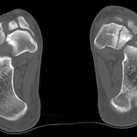
Weight-bearing foot radiographs demonstrated bilateral navicular deformities (comma-shaped) with possible fragmentation (Figures 1a, 1b and 1c). CT scan of the bilateral feet demonstrated a left navicular fracture with nonunion, in addition to confirming the radiographic findings (Figures 2a and 2b).
The patient underwent open surgery for the left navicular fracture, with bone graft placement and screw fixation. Post-surgical radiograph of the left foot showed appropriate internal fixation with the screw in place (Figure 3).
Discussion
Mueller–Weiss syndrome is a rare condition characterised by avascular necrosis of the tarsal navicular bone in the foot. It typically affects adults, with a higher prevalence in females aged between 40 to 60 years [1,2]. The exact cause remains unclear, but it is believed to involve a combination of vascular insufficiency, mechanical stress, and genetic factors [3]. The central third of the navicular is a watershed area between the medial plantar artery and the dorsalis pedis artery, and this area is prone to avascular necrosis and stress fractures [2]. Childhood-onset osteochondrosis of the navicular bone is called Köhler disease [2]. Note that Köhler disease would apply to paediatric patients, not adults.
Patients with Mueller–Weiss syndrome often present with insidious onset of foot pain, localised to the medial aspect of the midfoot. The pain is typically exacerbated by weight-bearing activities and may be associated with swelling. Clinical presentation and imaging studies can help differentiate Mueller–Weiss syndrome from other causes of midfoot pain, such as stress fractures, osteoarthritis, and other inflammatory conditions. While a fracture is seen in this case, the underlying pathology is Mueller–Weiss syndrome with bilateral navicular deformities.
Weight-bearing foot radiographs are the standard imaging method for diagnosis and disease staging. Radiographs may initially appear normal but can later reveal collapse and sclerosis of the navicular bone. A comma-shaped appearance of the navicular can be observed [2]. CT scan allows a more detailed evaluation of the extent of deformity, identifying arthritic changes and potential fractures. MRI can detect early signs of avascular necrosis, bone marrow oedema, and subchondral fracture lines. MRI also helps differentiate Mueller–Weiss syndrome from other conditions by assessing the extent of bone involvement and identifying associated soft tissue abnormalities.
Once the diagnosis of Mueller–Weiss syndrome is confirmed, treatment options can be considered. Non-surgical management, including activity modification, non-steroidal anti-inflammatory drugs (NSAIDs), and orthotic devices, are typically attempted initially [2]. In cases where conservative measures fail or in patients with navicular fracture nonunion, surgical intervention may be necessary, such as navicular bone decompression, internal fixation, bone grafting, or joint fusions [2,4].
The patient is doing well in follow-ups after the open surgery. He is able to walk and exercise with no pain and has gradually returned to normal activities. Understanding the typical clinical presentation and complications, the importance of imaging, and the available treatment options can aid in the accurate management of Mueller–Weiss syndrome.
All patient data have been completely anonymised throughout the entire manuscript and related files.
Differential Diagnosis List
Final Diagnosis
Mueller–Weiss syndrome (bilateral)
Liscense
This work is licensed under a Creative Commons Attribution-NonCommercial-ShareAlike 4.0 International License.
Figures
Standing AP & oblique radiographs of the feet


Axial & oblique axial CT images of the feet

Post-surgical radiograph of the left foot

1. Radiological Findings
Based on the provided bilateral foot X-rays and CT images, the following observations can be made:
- Bilateral navicular bones (especially on the left) show an abnormal shape, presenting a “comma-like” contour, with localized collapse and sclerosis.
- A noticeable fissure line is observed in the left navicular bone, suggesting a fracture (possibly due to repetitive stress or compromised blood supply).
- CT images further confirm collapse and deformation of the navicular bone, showing disorganized and sclerotic trabeculae, potential fracture lines, and irregular margins.
- No obvious significant narrowing of the joint spaces or marked soft tissue swelling is observed, but the surrounding joints of both navicular bones may be affected.
Overall imaging findings suggest bilateral navicular bone vascular compromise, collapse, and signs of stress concentration, with a visible fracture line on the left side.
2. Potential Diagnosis
Considering the patient’s age (23 years old), chronic bilateral foot pain exacerbated by activity, and the above imaging findings, the possible diagnoses include:
- Mueller–Weiss syndrome
This syndrome is a specific adult-onset avascular necrosis of the navicular bone, commonly characterized by navicular collapse, sclerosis, and a “comma-like” deformity on imaging. It differs from Köhler disease, which typically occurs in children. Given the patient’s current adult status and the characteristic imaging findings, Mueller–Weiss syndrome should be highly suspected. - Navicular Stress Fracture
A stress fracture usually results from excessive weight-bearing or repetitive microtrauma. It often appears as fracture lines along the medial or lateral cortex; however, isolated stress fractures typically do not present with pronounced avascular changes or collapse. Without clear evidence of vascular compromise, a simple stress fracture would be marked by localized fracture lines or periosteal reactions only. - Inflammatory Joint Disease (e.g., Rheumatoid Arthritis)
Such conditions can lead to multiple joint pains in the foot; however, they usually exhibit changes in joint space, soft tissue swelling, or erosive alterations, which are not fully consistent with the current imaging findings.
Considering the patient's age, clinical course, and radiological signs, Mueller–Weiss syndrome with associated navicular fracture is the most likely diagnosis.
3. Final Diagnosis
Taking into account the bilateral foot symptoms (pain worsened by weight-bearing and relieved by rest), the imaging findings (bilateral navicular “comma-like” deformity, sclerosis, local fracture), and ruling out other common causes of tarsal stress fractures or inflammatory pain, the most probable diagnosis is:
Mueller–Weiss syndrome (with bilateral navicular necrosis and collapse, and a fracture on the left side)
4. Treatment Plan and Rehabilitation
Based on the diagnosis of Mueller–Weiss syndrome and the patient’s current condition, the following treatment and rehabilitation options may be considered:
- Conservative Management
- Activity Modification: Reduce excessive weight-bearing on the affected feet. Avoid prolonged standing or walking. Gradually increase weight-bearing as advised by the physician.
- Medication: Non-steroidal anti-inflammatory drugs (NSAIDs) to alleviate pain and inflammation. If there is significant osteoporosis, consider calcium or vitamin D supplementation.
- Orthotic Devices or Special Insoles: Using arch supports and foot offloading can help reduce stress on the navicular area.
- Surgical Intervention
- Navicular Decompression, Internal Fixation, and Bone Grafting: Alleviates local pressure, promotes blood flow, and facilitates fracture healing.
- Arthrodesis: For severe joint deformities or significant joint surface destruction, fusion can stabilize the joint and relieve pain.
When conservative treatment is less effective or in cases of nonunion, surgery may be considered:
Rehabilitation/Exercise Prescription Recommendations (FITT-VP Principle):
- Frequency: Begin low-intensity foot exercises at home 2-3 times per week; increase gradually to 3-5 times per week as pain subsides and healing progresses.
- Intensity: Exercise should not trigger significant pain or discomfort. Start with seated or minimally weight-bearing foot movements.
- Time: Begin each session with 5-10 minutes, progressively extending to 15-20 minutes. You may break sessions into segments to avoid fatigue.
- Type:
- Range of Motion Exercises: Dorsiflexion and plantar flexion of the ankle, avoiding excessive force or speed.
- Resistance Band Training: Under professional guidance, perform progressive resistance exercises for the ankle and foot muscles.
- Low-Impact Aerobic Activities: Swimming or using a stationary bike can aid transitional weight-bearing once pain improves.
- Progression: As pain decreases and tolerance for weight-bearing increases, gradually lengthen walking intervals or intensity. Later on, introduce moderate-impact exercises such as jogging if tolerated.
Throughout rehabilitation, be vigilant for signs of excessive fatigue or reinjury. Should marked pain or swelling arise, seek medical attention and adjust the rehabilitation plan accordingly.
Note: If the patient has osteoporosis or other underlying conditions, further evaluation and risk assessment by specialized physicians may be necessary to tailor treatment and rehabilitation intensity.
Disclaimer: This report is solely a reference analysis based on the provided information and does not replace in-person consultation or the opinion of a qualified medical professional. If you have any concerns or experience changes in your condition, promptly seek advice from an orthopedic or relevant specialist for individualized recommendations.
Human Doctor Final Diagnosis
Mueller–Weiss syndrome (bilateral)