

A 70-year-old woman was admitted to the Emergency Department in 2014 after a standing height simple fall. She was diagnosed with a right subtrochanteric fracture and was treated with intramedullary nail fixation without complications. The patient had a medical history of diabetes mellitus, arterial hypertension, rheumatoid arthritis, depression and osteoporosis.
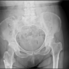
The initial anteroposterior radiograph of the pelvis and femur revealed a right subtrochanteric fracture with features consistent with an atypical fracture pattern. Also, there was a left focal subtrochanteric lateral cortical thickening not appreciated at the time.
A follow-up radiograph, 4 months after intramedullary nail fixation, showed delayed healing and left focal subtrochanteric lateral cortical thickening.
The patient was discharged from the clinic after 1 year, with the fracture consolidated and cortical thickening of the contralateral femur.
In 2016, she suffered an atypical complete subtrochanteric fracture of the left femur. The left-sided fracture was noted to run through the site of the previously demonstrated cortical beaking.
In 2016 the patient returned with pain and functional impotence of the left lower limb. The patient described "a crack sound" while walking alongside her husband but no mention of a fall. She was diagnosed with a complete subtrochanteric fracture of the left femur which was again managed with an intramedullary nail.
She had been on ibandronate for more than 10 years prior to her fractures. Long-term bisphosphonate therapy is associated with an increased risk of atypical femoral fractures (AFF). Early identification and intervention is essential to avoid the associated morbidity and mortality of a complete fracture [1].
In this case the left femoral findings were overlooked, leading to a delay in diagnosis and treatment.
The American Society for Bone and Mineral Research (ASBMR) Task Force revised the case definition of AFF in 2013. To satisfy the case definition of AFF, the fracture must be located along the femoral diaphysis from just distal to the lesser trochanter to just proximal to the suprachondylar flare. In addition, at least four of five major features must be present. None of the minor features is required but have been sometimes associated with these fractures [2].
This case suits the definition of an AFF. All of the major features are present: the fracture is associated with minimal or no trauma; the fracture line originates at the lateral cortex, is transverse in its orientation and becomes oblique as it progresses medially across the femur; the complete fracture extends through both cortices and is associated with a medial spike; the fracture is noncomminuted; there is a localised thickening of the lateral cortex at the fracture site. Minor features like generalised increase in cortical thickness of the diaphysis, delayed fracture healing and bilateral complete femoral diaphysis fractures are present.
According to the revised AFF case definition, features linking AFFs to comorbid conditions (diabetes, rheumatoid arthritis, vitamin D deficiency) and drugs (ie. bisphosphonates, glucocorticoids, proton pump inhibitors), were removed from the 2010 criteria, and deemed more appropriate for studies of associations [2].
In this case, according to the previous definition the patient had several risk factors, so we cannot conclude that bisphosphonates are the only possible culprit for this event. Nonetheless, they are the most studied and represented in the literature.
Written informed patient consent for publication has been obtained.
Bilateral atypical femoral fracture
This work is licensed under a Creative Commons Attribution-NonCommercial-ShareAlike 4.0 International License.


Based on the provided X-ray images of the pelvis and bilateral femurs:
Overall imaging findings suggest that the left femur exhibits a typical subhip transverse-to-oblique fracture trajectory with lateral cortical thickening, consistent with the characteristics of an Atypical Femoral Fracture (AFF).
Based on the patient’s history (over 10 years of bisphosphonate use, bilateral femoral fractures in similar locations) and imaging findings, possible diagnoses or differential diagnoses include:
Based on:
Overall, the most likely diagnosis is: Atypical Femoral Fracture (AFF).
If uncertainty remains, MRI or bone scintigraphy can be performed to rule out potential neoplastic processes, and bone metabolic markers or laboratory tests can be used to comprehensively assess bone turnover status.
During rehabilitation, it is crucial to prevent further falls or fractures; seek guidance from a professional rehabilitation therapist if necessary.
Disclaimer: This report is a reference analysis based on existing information and does not replace an in-person consultation or professional orthopedic guidance. If you have any doubts or experience worsening symptoms, please seek medical attention promptly.
Bilateral atypical femoral fracture