


A 68-year-old male patient with known hypertension, dyslipemia, auricular fibrillation and peripheral vasculopathy, presented with left sciatic pain and non-symptomatic hips.
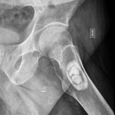
Radiographs showed a bone tumour, in the centromedular intertrochanteric region of the left femur (Figs 1 and 2). It has well-defined limits, doubtful chondroid matrix and absence of aggressive signs (Fig 3). CT confirmed the presence of a geographic hypodense lesion with sclerotic borders and dystrophic internal mineralised matrix (Fig 4). On MRI (Figs 5, 6 7), the non-calcified component was hypointense to muscle on T1 and very hyperintense on T2-FATSAT. Note is made on a small fatty nodule in the non-calcified tumoural matrix.
Liposclerosing myxofibrous tumour (LSMFT) is a rare fibro-osseous benign tumour [2]. It is formed by the combination of several elements with variable proportions: lipoma, fibroxanthoma, myxoma, myxofibroma, fibrous tissue, cyst formation, fat necrosis, ischaemic bone tissue, and rarely cartilaginous tissue [1]. LSMFT affects men and women equally in the fourth decade of life [3], without ethnical predilection [2]. Histological and genetic analysis suggested that it may represent a variant or evolution of fibrous dysplasia or intraosseous lipoma, probably because of stress or fatigue suffered by the initial lesion [2].
The frequent presentation is an incidental finding in asymptomatic patients. In cases of being symptomatic, pain and pathologic fractures are the most common presentations[2].
Bone biopsy may be hard to interpret and the results may be confounding because of the heterogeneity of the tumour matrix. Nowadays one is discouraged unless with imaging demonstration of aggressive features [2].
The radiologic appearance and the skeletal distribution of LSMFT are typical and establish the diagnosis. It has an important predilection for the femur (85%) and for the intertrochanteric region (91%) [1, 2]. CT and MRI show a well-defined geographic lesion, often with a thick and extensive sclerotic margin, reflecting its slow growing pattern, sometimes with globular and irregular mineralised matrix. After venous contrast administration, the myxoid matrix may show some enhancement.
MR and/or CT are essential to differentiate from intraosseous lipoma because they offer the possibility of documenting intralesional fat, which is minimal if any in LSMFT. Fatty areas demonstrate low attenuation values (<-60HU) on CT, high SI on T1/T2WI and low SI on FS/STIR on MRI. The differentiation from intraosseous lipoma with involutional change may not be possible through imaging studies but the intertrochanteric location favours the diagnosis of LSMFT [2].
Fibrous dysplasia (FD) reveals less sclerosis, usually with the characteristic “ground glass” pattern and endosteal scalloping, less frequent in LSMFT [2].
In bone scintigraphy both FD and LSMFT may show low tracer uptake [3].
Malignant transformation to osteosarcoma or malignant fibrous histiocytoma occurs approximately in 10-16% [1]. It is suggested in imaging when aggressive signs are developed, such as cortical disruption, rapid growing and soft-tissue extension. Treatment is considered in symptomatic patients, which includes curettage, bone-grafting and fixation. Cases with malignant transformations or pathological fracture, usually require arthroplasty [3]. The non-surgical treatment includes analgesics and follow-up with plain X-ray every six months and MR or CT every year during the first two years [2].
Radiology can lead to the diagnosis.
Histology can cause confusion.
Written informed patient consent for publication has been obtained.
Liposclerosing myxofibrous tumour (LSMFT)
This work is licensed under a Creative Commons Attribution-NonCommercial-ShareAlike 4.0 International License.


The patient is a 68-year-old male with a history of hypertension, dyslipidemia, atrial fibrillation, and peripheral vascular disease, presenting with left-sided sciatic pain. Imaging examinations (including X-ray, CT, and MRI) targeting the left hip and proximal femur reveal:
Based on the above imaging findings and the patient’s history, the possible diagnoses or differentials include:
Considering the patient’s age, symptoms (relatively mild pain), and imaging features (a lesion in the proximal femur intertrochanteric zone with a thick sclerotic margin, mixed matrix, and no prominent fat signal), the most likely diagnosis is Liposclerosing Myxofibrous Tumor (LSMFT).
For further confirmation, imaging follow-up can be performed. If suspicious malignant features (such as rapid lesion enlargement, cortical bone destruction, or soft tissue mass) appear, a biopsy may be indicated to rule out malignancy.
Liposclerosing myxofibrous tumor is usually a slowly growing benign lesion. Some cases can be managed conservatively under imaging surveillance. Key points in treatment and rehabilitation include:
Disclaimer: This report provides a reference analysis based on the available imaging and clinical information. It does not replace an in-person consultation or professional diagnosis and treatment. If you have any questions or changes in your condition, please consult an orthopedic or related specialist promptly.
Liposclerosing myxofibrous tumour (LSMFT)