


A previously healthy 9-year-old child was admitted to hospital with severe post-traumatic left-hand pain. Physical examination showed slight swelling on the hand back, without bruising or deformity. A hard consistency tumour was identified under palpation of the back of the second metacarpal. Mobility of fingers and wrist was preserved.
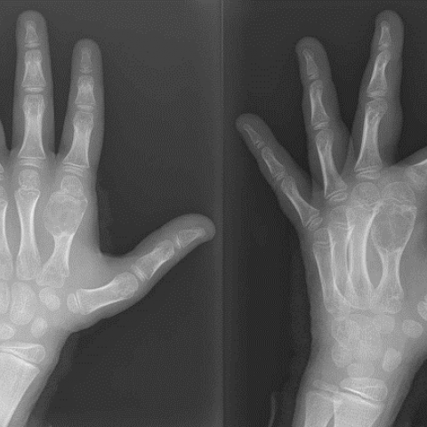
Emergency left-hand X-ray showed a mostly lithic metaphyseal-diaphyseal bone lesion in second metacarpal (Figure. 1). The lesion presented cortical thinning as well as ridges and partitions inside, without periosteal reaction.
Left hand MRI confirmed the lesion (Figure. 2), which showed hypointense signal in T2-weighted sequences (Figure. 2 A), hyperintense signal in PDWI (Figure. 2 B) and STIR (Figure. 2 D), and intermediate signal in T1-weighted sequences (Figure. 2 C). The lesion also displayed hypointense punctate focus inside. It extended through the metaphysis and reached the epiphysis, with the presence of a slight soft tissue component at flexor aspect. Intense diffuse postcontrast enhancement was maintained in time (Figure. 2 E).
First differential diagnosis reported either a giant cell tumour or a giant cell reparative granuloma. Incisional biopsy showed multinucleated osteoclasts with variable cytological atypia and mitotic activity, suggesting either an aggressive giant cell bone tumour or a giant-cell rich variant osteosarcoma.
Body-CT and bone scintigraphy were negative. Accordingly, patient underwent resection plus reconstruction of second metacarpal with bicortical iliac graft (Figure. 3). Osteotomy piece showed a conventional high-grade osteosarcoma, giant cell-rich variant (Figure. 4 and 5), without MDM2(12q15) amplification. Therefore, patient underwent neoadjuvant chemotherapy followed by amputation of second metacarpal and phalanges plus reconstruction using peroneal bone. Histological analysis confirmed absence of tumour cells.
Background:
Giant cell-rich osteosarcoma (GCRO) is an uncommon variant that accounts for 1-3% of all cases of conventional osteosarcomas. Cytologically shows malignant cells associated with osteoid deposition. Abundant admixed benign osteoclast-like giant cells may obscure malignant features [1].
Clinical Perspective:
GCRO usually appears in metaphyseal/metadiaphyseal location in a skeletally immature patient [1]. Most patients suffer from swelling and pain in the involved region, which tend to worsen over time and can cause limp when bearing weight. Systemic symptoms are very uncommon [2,3,4].
Imaging Perspective:
X-Ray usually demonstrates a mixed pattern of sclerosis and lucent areas, as well as characteristics of osteoid matrix production (variable amount of fluffy, cloudlike opacities). High-grade variants can show aggressive periosteal reaction (Codman triangle, laminated, hair-on-end or sunburst patterns) and soft tissue component [5-10]. Under MRI, GCRO usually shows areas of intermediate signal intensity on T1-WI and hyperintense areas replacing the normal marrow on T2-WI. Areas of low signal intensity on both T1-WI and T2-WI represent mineralised matrix. Central haemorrhage (high signal intensity in all sequences) and necrosis (low signal intensity on T1-WI and high signal intensity on T2-WI) are frequent. Extension through the open physis into the epiphysis is common. Fat-suppressed T1-WI gadolinium-enhanced images are helpful for delineating extension into the joint, although the synovium is rarely violated [10].
Outcome:
Treatment requires aggressive surgical resection, often with amputation, followed by chemotherapy [3,9]. If a limb-salvage procedure is feasible (80%–90% of patients), neoadjuvant chemotherapy precedes surgery to downstage the tumour [9,11-13]. Radiotherapy may be useful in patients who are unable to undergo complete resection or who have microscopic residual tumour foci [9,11].
Take-Home Message / Teaching Points
GCRO must be included in the differential diagnosis of primary giant-cell rich lesions of bone with metaphyseal-diaphyseal location in skeletally immature patients.
Giant cell-rich osteosarcoma
This work is licensed under a Creative Commons Attribution-NonCommercial-ShareAlike 4.0 International License.


According to the provided anteroposterior X-ray of the hand and MRI images, the following findings are noted:
Considering the patient's age of 9, severe local pain following trauma, the presence of mixed lytic and sclerotic changes on imaging, and tumor cells rich in multinucleated giant cells, the following diagnoses merit particular attention:
Taking into account the patient's age, clinical symptoms, imaging findings, and final pathological examination results (including multinucleated giant cells, definitive malignant components, osteoid formation, and the absence of MDM2(12q15) gene amplification consistent with high-grade osteosarcoma), the most likely final diagnosis is:
Giant Cell-Rich High-Grade Conventional Osteosarcoma
1. Treatment Strategy:
2. Rehabilitation and Exercise Prescription:
Disclaimer: This report is a reference analysis based on the information provided and should not replace an in-person clinical diagnosis or professional medical advice. If you have any questions or if your condition changes, please consult orthopedic and oncology specialists for further evaluation.
Giant cell-rich osteosarcoma